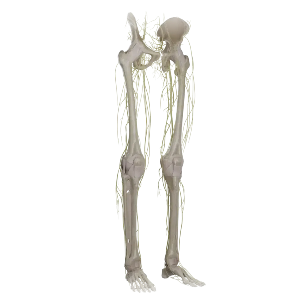

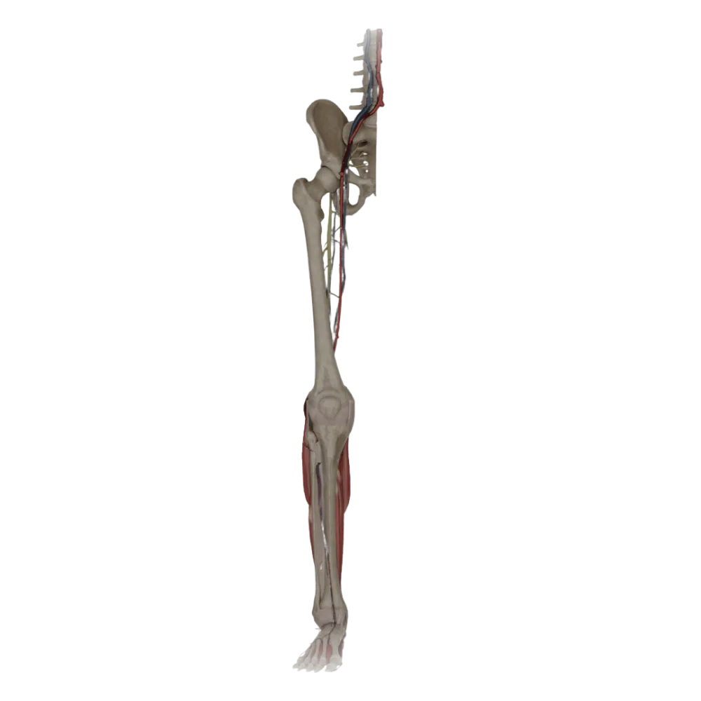
Test of the anatomy of the tibial nerve.
Evaluate your knowledge of the anatomy of the tibial nerve. This test strictly assesses its topography, branching, syntopy, and zones of innervation in the shin and foot.
1/20
bold
text
1. Where does the tibial nerve originate?
-
From the lumbar plexus.
The tibial nerve is the largest branch of the sciatic nerve, which itself is formed from the sacral plexus.
-
Is a branch of the femoral nerve.
The tibial nerve is the largest branch of the sciatic nerve, which itself is formed from the sacral plexus.
-
From the sacral plexus as an extension of the sciatic nerve.
The tibial nerve is the largest branch of the sciatic nerve, which itself is formed from the sacral plexus.
-
From the obturator nerve.
The tibial nerve is the largest branch of the sciatic nerve, which itself is formed from the sacral plexus.
-
I find it difficult to answer
The tibial nerve is the largest branch of the sciatic nerve, which itself is formed from the sacral plexus.
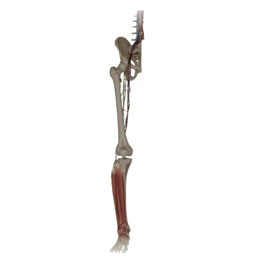
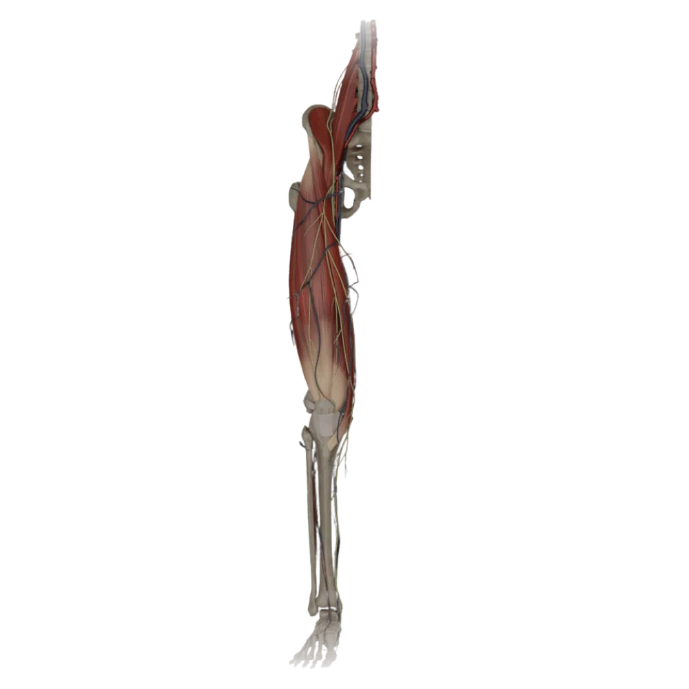
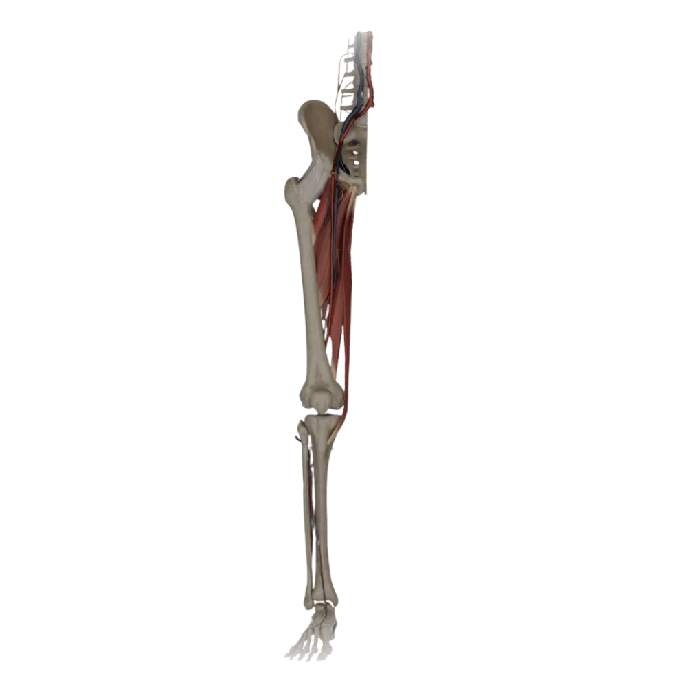
2. What is the syntopic position of the tibial nerve in the popliteal fossa relative to the popliteal vein and artery?
-
It lies most superficially and laterally.
In the popliteal fossa, structures are arranged (from surface inward and medially): nerve, vein, artery (NEVA rule). Thus, the nerve lies more superficial and lateral.
-
It lies the deepest and medially.
In the popliteal fossa, structures are arranged (from surface inward and medially): nerve, vein, artery (NEVA rule). Thus, the nerve lies more superficial and lateral.
-
It is located between the vein and the artery.
In the popliteal fossa, structures are arranged (from surface inward and medially): nerve, vein, artery (NEVA rule). Thus, the nerve lies more superficial and lateral.
-
Located medially to the artery.
In the popliteal fossa, structures are arranged (from surface inward and medially): nerve, vein, artery (NEVA rule). Thus, the nerve lies more superficial and lateral.
-
I find it difficult to answer
In the popliteal fossa, structures are arranged (from surface inward and medially): nerve, vein, artery (NEVA rule). Thus, the nerve lies more superficial and lateral.
3. Which canal does the tibial nerve enter when leaving the popliteal fossa?
-
Femoral canal (canalis femoralis).
After passing under the tendinous arch of the soleus muscle, the nerve, together with the posterior tibial artery and veins, enters the tarsal tunnel (Gruber's canal).
-
Adductor canal (canalis adductorius).
After passing under the tendinous arch of the soleus muscle, the nerve, together with the posterior tibial artery and veins, enters the tarsal tunnel (Gruber's canal).
-
Obturator canal (canalis obturatorius).
After passing under the tendinous arch of the soleus muscle, the nerve, together with the posterior tibial artery and veins, enters the tarsal tunnel (Gruber's canal).
-
Cruripoplietal canal (canalis cruropopliteus).
After passing under the tendinous arch of the soleus muscle, the nerve, together with the posterior tibial artery and veins, enters the tarsal tunnel (Gruber's canal).
-
I find it difficult to answer
After passing under the tendinous arch of the soleus muscle, the nerve, together with the posterior tibial artery and veins, enters the tarsal tunnel (Gruber's canal).
4. Which of the listed lower leg muscles is NOT innervated by the tibial nerve?
-
Tibialis posterior muscle.
The long fibular muscle belongs to the lateral group of lower leg muscles and is innervated by the superficial fibular nerve, not the tibial.
-
Fibularis longus muscle.
The long fibular muscle belongs to the lateral group of lower leg muscles and is innervated by the superficial fibular nerve, not the tibial.
-
Flexor digitorum longus
The long fibular muscle belongs to the lateral group of lower leg muscles and is innervated by the superficial fibular nerve, not the tibial.
-
Soleus muscle.
The long fibular muscle belongs to the lateral group of lower leg muscles and is innervated by the superficial fibular nerve, not the tibial.
-
I find it difficult to answer
The long fibular muscle belongs to the lateral group of lower leg muscles and is innervated by the superficial fibular nerve, not the tibial.
5. Which cutaneous branch does the tibial nerve in the popliteal fossa give off?
-
Lateral sural cutaneous nerve
In the popliteal fossa, the tibial nerve gives off the medial sural cutaneous nerve (n. cutaneus surae medialis), which descends along the posterior surface of the lower leg.
-
Medial sural cutaneous nerve.
In the popliteal fossa, the tibial nerve gives off the medial sural cutaneous nerve (n. cutaneus surae medialis), which descends along the posterior surface of the lower leg.
-
Saphenous nerve.
In the popliteal fossa, the tibial nerve gives off the medial sural cutaneous nerve (n. cutaneus surae medialis), which descends along the posterior surface of the lower leg.
-
Posterior femoral cutaneous nerve.
In the popliteal fossa, the tibial nerve gives off the medial sural cutaneous nerve (n. cutaneus surae medialis), which descends along the posterior surface of the lower leg.
-
I find it difficult to answer
In the popliteal fossa, the tibial nerve gives off the medial sural cutaneous nerve (n. cutaneus surae medialis), which descends along the posterior surface of the lower leg.
6. By the fusion of which nerves is the sural nerve (n. suralis) formed?
-
Of the saphenous nerve and the medial sural cutaneous nerve.
The sural nerve is formed in the lower third of the leg by the union of the medial sural cutaneous nerve (from the tibial) and the lateral sural cutaneous nerve (from the common fibular).
-
Of the superficial fibular and lateral cutaneous nerves.
The sural nerve is formed in the lower third of the leg by the union of the medial sural cutaneous nerve (from the tibial) and the lateral sural cutaneous nerve (from the common fibular).
-
Of the medial and lateral sural cutaneous nerves.
The sural nerve is formed in the lower third of the leg by the union of the medial sural cutaneous nerve (from the tibial) and the lateral sural cutaneous nerve (from the common fibular).
-
Of the deep fibular and saphenous nerves.
The sural nerve is formed in the lower third of the leg by the union of the medial sural cutaneous nerve (from the tibial) and the lateral sural cutaneous nerve (from the common fibular).
-
I find it difficult to answer
The sural nerve is formed in the lower third of the leg by the union of the medial sural cutaneous nerve (from the tibial) and the lateral sural cutaneous nerve (from the common fibular).
7. In what order (anterior to posterior) are the structures located behind the medial malleolus?
-
Artery, vein, nerve, tendon of m. tibialis posterior
Behind the medial malleolus, structures run anterior to posterior: tendons of m. tibialis posterior and m. flexor digitorum longus, then vessels and nerve, and posteriorly tendon of m. flexor hallucis longus
-
N. tibialis, artery and vein, flexor tendons.
Behind the medial malleolus, structures run anterior to posterior: tendons of m. tibialis posterior and m. flexor digitorum longus, then vessels and nerve, and posteriorly tendon of m. flexor hallucis longus
-
Tendon m. flexor hallucis longus, n. tibialis, artery and vein, tendon m. flexor digitorum longus
Behind the medial malleolus, structures run anterior to posterior: tendons of m. tibialis posterior and m. flexor digitorum longus, then vessels and nerve, and posteriorly tendon of m. flexor hallucis longus
-
Tendon m. tibialis posterior, tendon m. flexor digitorum longus, posterior tibial artery and vein, n. tibialis, tendon m. flexor hallucis longus
Behind the medial malleolus, structures run anterior to posterior: tendons of m. tibialis posterior and m. flexor digitorum longus, then vessels and nerve, and posteriorly tendon of m. flexor hallucis longus
-
I find it difficult to answer
Behind the medial malleolus, structures run anterior to posterior: tendons of m. tibialis posterior and m. flexor digitorum longus, then vessels and nerve, and posteriorly tendon of m. flexor hallucis longus
8. Into which terminal branches does the tibial nerve divide?
-
Superficial and deep fibular nerves.
After passing beneath the flexor retinaculum, the tibial nerve divides into its terminal branches: medial and lateral plantar nerves.
-
Medial and lateral plantar nerves.
After passing beneath the flexor retinaculum, the tibial nerve divides into its terminal branches: medial and lateral plantar nerves.
-
Medial and lateral calcaneal nerves.
After passing beneath the flexor retinaculum, the tibial nerve divides into its terminal branches: medial and lateral plantar nerves.
-
Dorsal and plantar nerves of the foot.
After passing beneath the flexor retinaculum, the tibial nerve divides into its terminal branches: medial and lateral plantar nerves.
-
I find it difficult to answer
After passing beneath the flexor retinaculum, the tibial nerve divides into its terminal branches: medial and lateral plantar nerves.
9. Which muscle is innervated by the medial plantar nerve?
-
Flexor digitorum brevis muscle.
The medial plantar nerve innervates the abductor hallucis muscle, flexor digitorum brevis, flexor hallucis brevis, and the first lumbrical muscle.
-
Abductor digiti minimi muscle.
The medial plantar nerve innervates the abductor hallucis muscle, flexor digitorum brevis, flexor hallucis brevis, and the first lumbrical muscle.
-
Quadratus plantae muscle.
The medial plantar nerve innervates the abductor hallucis muscle, flexor digitorum brevis, flexor hallucis brevis, and the first lumbrical muscle.
-
The muscle adducting the great toe of the foot.
The medial plantar nerve innervates the abductor hallucis muscle, flexor digitorum brevis, flexor hallucis brevis, and the first lumbrical muscle.
-
I find it difficult to answer
The medial plantar nerve innervates the abductor hallucis muscle, flexor digitorum brevis, flexor hallucis brevis, and the first lumbrical muscle.
10. What is the zone of sensory innervation of the medial plantar nerve on the foot?
-
The skin of the lateral one and a half toes and the corresponding part of the sole.
The sensory branches of the medial plantar nerve provide innervation to the skin of the medial part of the sole and the plantar surface of the first 3.5 toes.
-
The skin of the dorsum of the foot.
The sensory branches of the medial plantar nerve provide innervation to the skin of the medial part of the sole and the plantar surface of the first 3.5 toes.
-
The skin of the heel region.
The sensory branches of the medial plantar nerve provide innervation to the skin of the medial part of the sole and the plantar surface of the first 3.5 toes.
-
The skin of the medial three and a half toes and corresponding part of the sole.
The sensory branches of the medial plantar nerve provide innervation to the skin of the medial part of the sole and the plantar surface of the first 3.5 toes.
-
I find it difficult to answer
The sensory branches of the medial plantar nerve provide innervation to the skin of the medial part of the sole and the plantar surface of the first 3.5 toes.
11. Which muscle is innervated by the deep branch of the lateral plantar nerve?
-
Flexor digitorum brevis muscle.
The deep branch of the lateral plantar nerve supplies the adductor hallucis muscle, interosseous muscles, and lateral lumbrical muscles.
-
Abductor hallucis muscle.
The deep branch of the lateral plantar nerve supplies the adductor hallucis muscle, interosseous muscles, and lateral lumbrical muscles.
-
The muscle adducting the great toe of the foot.
The deep branch of the lateral plantar nerve supplies the adductor hallucis muscle, interosseous muscles, and lateral lumbrical muscles.
-
First lumbrical muscle
The deep branch of the lateral plantar nerve supplies the adductor hallucis muscle, interosseous muscles, and lateral lumbrical muscles.
-
I find it difficult to answer
The deep branch of the lateral plantar nerve supplies the adductor hallucis muscle, interosseous muscles, and lateral lumbrical muscles.
12. Through which anatomical structure does the tibial nerve pass to the foot?
-
Beneath the flexor retinaculum (retinaculum musculorum flexorum).
The nerve passes behind the medial malleolus in the osteofibrous canal under the flexor retinaculum, after which it emerges onto the sole.
-
Through the inferior muscular-peroneal canal.
The nerve passes behind the medial malleolus in the osteofibrous canal under the flexor retinaculum, after which it emerges onto the sole.
-
Beneath the lateral malleolus.
The nerve passes behind the medial malleolus in the osteofibrous canal under the flexor retinaculum, after which it emerges onto the sole.
-
Through the interosseous membrane of the leg.
The nerve passes behind the medial malleolus in the osteofibrous canal under the flexor retinaculum, after which it emerges onto the sole.
-
I find it difficult to answer
The nerve passes behind the medial malleolus in the osteofibrous canal under the flexor retinaculum, after which it emerges onto the sole.
13. Which nerve is the medial calcaneal branches (rr. calcanei mediales) a branch of?
-
Sural nerve.
The medial calcaneal branches directly branch from the trunk of the tibial nerve (before its division into plantar nerves) and pierce the flexor retinaculum.
-
Deep fibular nerve.
The medial calcaneal branches directly branch from the trunk of the tibial nerve (before its division into plantar nerves) and pierce the flexor retinaculum.
-
Lateral plantar nerve.
The medial calcaneal branches directly branch from the trunk of the tibial nerve (before its division into plantar nerves) and pierce the flexor retinaculum.
-
Tibial nerve
The medial calcaneal branches directly branch from the trunk of the tibial nerve (before its division into plantar nerves) and pierce the flexor retinaculum.
-
I find it difficult to answer
The medial calcaneal branches directly branch from the trunk of the tibial nerve (before its division into plantar nerves) and pierce the flexor retinaculum.
14. Which of the following statements about the syntopy of the lateral plantar nerve is correct?
-
It passes between the long and short flexors of the toes.
The lateral plantar nerve moves out and forward, passing between the flexor digitorum brevis and the quadratus plantae.
-
It is located between the flexor digitorum brevis and the quadratus plantae.
The lateral plantar nerve moves out and forward, passing between the flexor digitorum brevis and the quadratus plantae.
-
It travels in the medial plantar groove.
The lateral plantar nerve moves out and forward, passing between the flexor digitorum brevis and the quadratus plantae.
-
It lies superficial to the plantar aponeurosis.
The lateral plantar nerve moves out and forward, passing between the flexor digitorum brevis and the quadratus plantae.
-
I find it difficult to answer
The lateral plantar nerve moves out and forward, passing between the flexor digitorum brevis and the quadratus plantae.
15. Which lumbrical muscles of the foot are innervated by the lateral plantar nerve?
-
Only the first
The lateral plantar nerve innervates the 2nd, 3rd, and 4th lumbrical muscles, whereas the 1st is innervated by the medial plantar nerve.
-
First and second
The lateral plantar nerve innervates the 2nd, 3rd, and 4th lumbrical muscles, whereas the 1st is innervated by the medial plantar nerve.
-
The second, third, and fourth.
The lateral plantar nerve innervates the 2nd, 3rd, and 4th lumbrical muscles, whereas the 1st is innervated by the medial plantar nerve.
-
All lumbrical muscles
The lateral plantar nerve innervates the 2nd, 3rd, and 4th lumbrical muscles, whereas the 1st is innervated by the medial plantar nerve.
-
I find it difficult to answer
The lateral plantar nerve innervates the 2nd, 3rd, and 4th lumbrical muscles, whereas the 1st is innervated by the medial plantar nerve.
16. Which of the lower leg muscles receives innervation from the tibial nerve before it enters the tarsal tunnel?
-
Popliteal muscle.
The popliteus muscle, located on the posterior surface of the knee joint capsule, receives branches from the tibial nerve directly in the popliteal fossa.
-
Extensor digitorum longus.
The popliteus muscle, located on the posterior surface of the knee joint capsule, receives branches from the tibial nerve directly in the popliteal fossa.
-
Fibularis brevis muscle.
The popliteus muscle, located on the posterior surface of the knee joint capsule, receives branches from the tibial nerve directly in the popliteal fossa.
-
Fibularis tertius muscle.
The popliteus muscle, located on the posterior surface of the knee joint capsule, receives branches from the tibial nerve directly in the popliteal fossa.
-
I find it difficult to answer
The popliteus muscle, located on the posterior surface of the knee joint capsule, receives branches from the tibial nerve directly in the popliteal fossa.
17. Where is the tibial nerve topographically located in the tarsal tunnel?
-
Between the anterior tibial muscle and the interosseous membrane.
The popliteal canal is formed anteriorly by the deep muscles of the posterior group (m. tibialis posterior and others) and posteriorly by the soleus muscle (superficial layer).
-
Between the superficial and deep muscles of the posterior lower leg group.
The popliteal canal is formed anteriorly by the deep muscles of the posterior group (m. tibialis posterior and others) and posteriorly by the soleus muscle (superficial layer).
-
In the thickness of the medial head of the gastrocnemius muscle.
The popliteal canal is formed anteriorly by the deep muscles of the posterior group (m. tibialis posterior and others) and posteriorly by the soleus muscle (superficial layer).
-
Between the fibula and the long fibular muscle.
The popliteal canal is formed anteriorly by the deep muscles of the posterior group (m. tibialis posterior and others) and posteriorly by the soleus muscle (superficial layer).
-
I find it difficult to answer
The popliteal canal is formed anteriorly by the deep muscles of the posterior group (m. tibialis posterior and others) and posteriorly by the soleus muscle (superficial layer).
18. Which artery accompanies the tibial nerve in the popliteal canal?
-
Anterior tibial artery.
In the popliteal canal, the tibial nerve runs as part of the neurovascular bundle along with the posterior tibial artery and its accompanying veins.
-
Popliteal artery.
In the popliteal canal, the tibial nerve runs as part of the neurovascular bundle along with the posterior tibial artery and its accompanying veins.
-
Fibular artery
In the popliteal canal, the tibial nerve runs as part of the neurovascular bundle along with the posterior tibial artery and its accompanying veins.
-
Posterior tibial artery.
In the popliteal canal, the tibial nerve runs as part of the neurovascular bundle along with the posterior tibial artery and its accompanying veins.
-
I find it difficult to answer
In the popliteal canal, the tibial nerve runs as part of the neurovascular bundle along with the posterior tibial artery and its accompanying veins.
19. Which muscle of the foot is innervated by the lateral plantar nerve?
-
Flexor hallucis brevis
Among the listed muscles, only the quadratus plantae receives innervation from the lateral plantar nerve. The rest are innervated by the medial plantar nerve.
-
Abductor hallucis muscle.
Among the listed muscles, only the quadratus plantae receives innervation from the lateral plantar nerve. The rest are innervated by the medial plantar nerve.
-
Quadratus plantae muscle.
Among the listed muscles, only the quadratus plantae receives innervation from the lateral plantar nerve. The rest are innervated by the medial plantar nerve.
-
First lumbrical muscle
Among the listed muscles, only the quadratus plantae receives innervation from the lateral plantar nerve. The rest are innervated by the medial plantar nerve.
-
I find it difficult to answer
Among the listed muscles, only the quadratus plantae receives innervation from the lateral plantar nerve. The rest are innervated by the medial plantar nerve.
20. Which joints are innervated by the articular branches of the tibial nerve?
-
Knee and ankle
The articular branches of the tibial nerve participate in innervation of the knee joint capsule (in the popliteal fossa) and the ankle joint (in the medial malleolus area).
-
Hip and knee
The articular branches of the tibial nerve participate in innervation of the knee joint capsule (in the popliteal fossa) and the ankle joint (in the medial malleolus area).
-
Only joints of the foot
The articular branches of the tibial nerve participate in innervation of the knee joint capsule (in the popliteal fossa) and the ankle joint (in the medial malleolus area).
-
Sacroiliac and hip
The articular branches of the tibial nerve participate in innervation of the knee joint capsule (in the popliteal fossa) and the ankle joint (in the medial malleolus area).
-
I find it difficult to answer
The articular branches of the tibial nerve participate in innervation of the knee joint capsule (in the popliteal fossa) and the ankle joint (in the medial malleolus area).
Retake this quiz?
Your current progress will be reset.