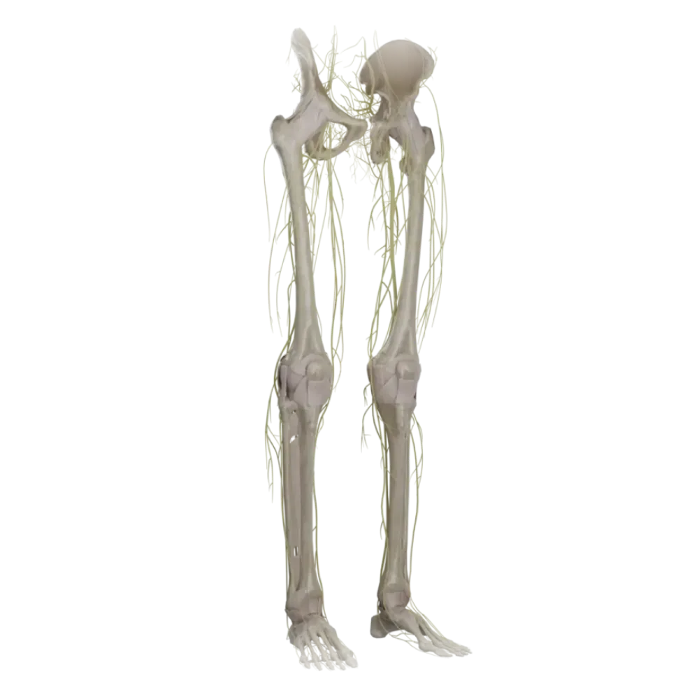
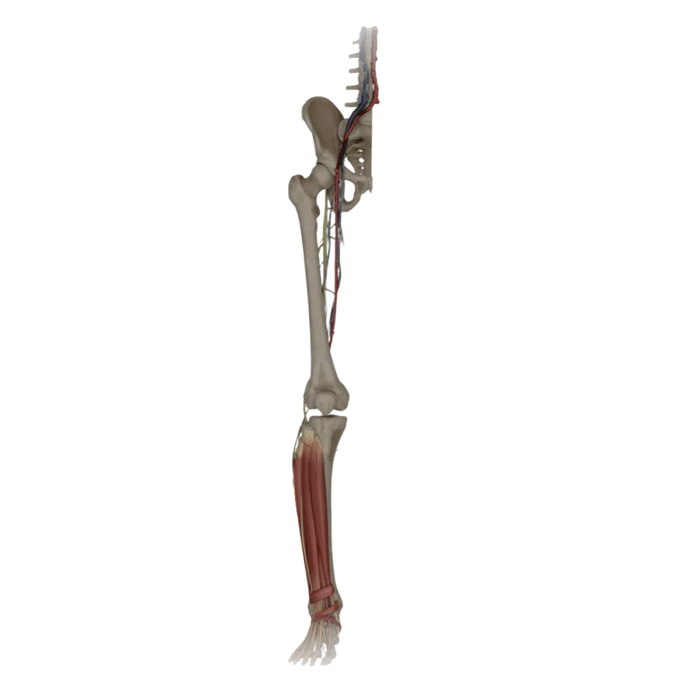
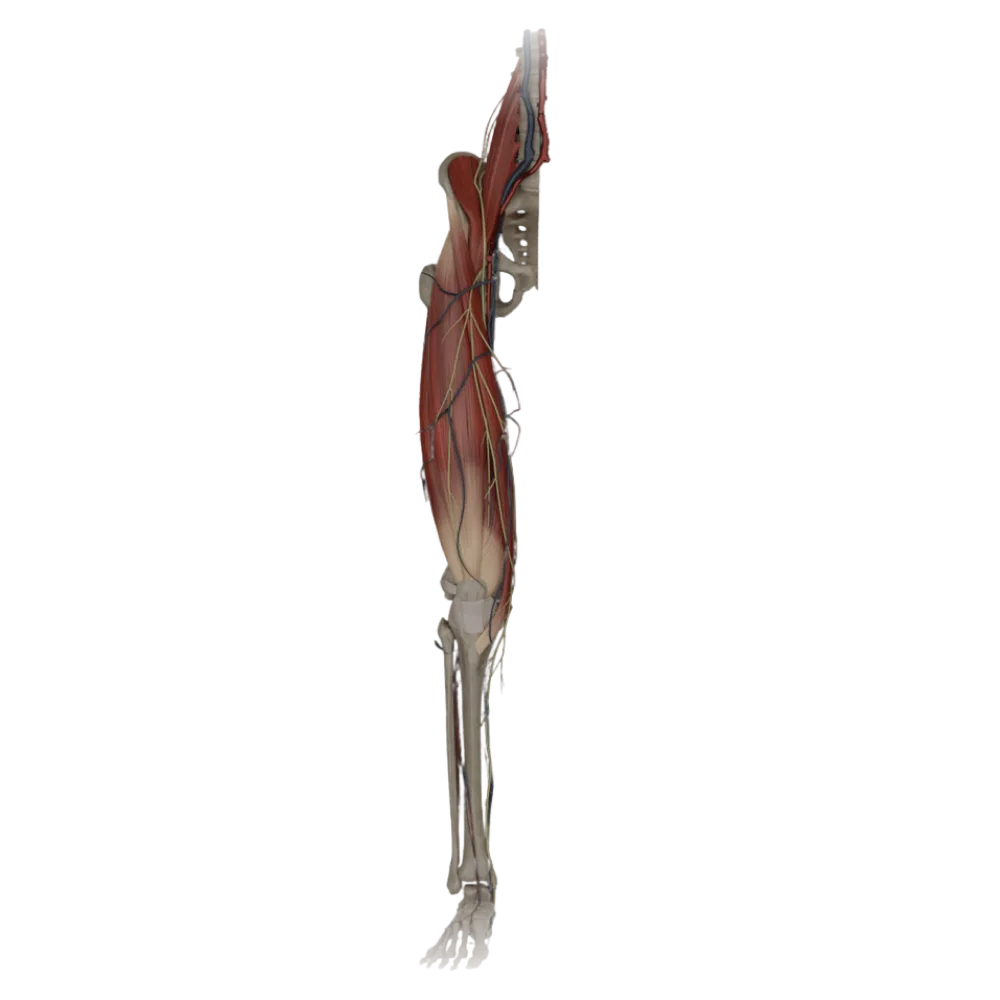
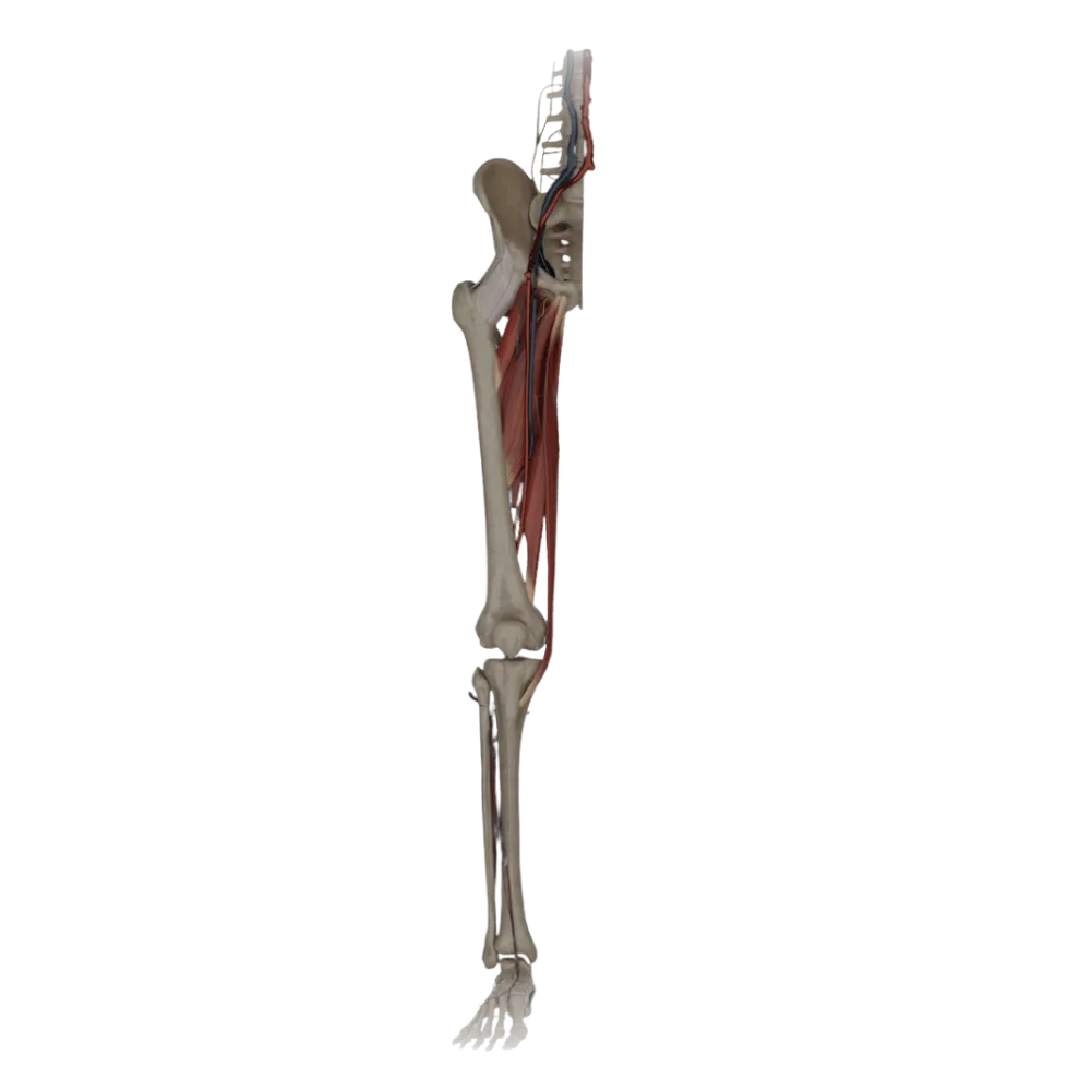

Femoral nerve anatomy test
Evaluate the knowledge of femoral nerve anatomy. The test thoroughly examines its topography, muscular and sensory branches, as well as innervation zones.
1/20
bold
text
1. From which segments of the spinal cord is the femoral nerve formed?
-
L1-L2
The femoral nerve is the largest branch of the lumbar plexus and is formed from the anterior rami of the L2-L4 spinal nerves.
-
L2 - L4
The femoral nerve is the largest branch of the lumbar plexus and is formed from the anterior rami of the L2-L4 spinal nerves.
-
L4 - S3
The femoral nerve is the largest branch of the lumbar plexus and is formed from the anterior rami of the L2-L4 spinal nerves.
-
S1-S4
The femoral nerve is the largest branch of the lumbar plexus and is formed from the anterior rami of the L2-L4 spinal nerves.
-
I find it difficult to answer
The femoral nerve is the largest branch of the lumbar plexus and is formed from the anterior rami of the L2-L4 spinal nerves.
2. Along which border of the psoas major muscle (m. psoas major) does the femoral nerve trunk travel in the abdominal cavity?
-
Along the lateral border
The nerve lies in the groove between the iliacus and psoas major, emerging from under the lateral border of the latter.
-
Along the medial border
The nerve lies in the groove between the iliacus and psoas major, emerging from under the lateral border of the latter.
-
On the anterior surface
The nerve lies in the groove between the iliacus and psoas major, emerging from under the lateral border of the latter.
-
Pierces the thickness of the muscle
The nerve lies in the groove between the iliacus and psoas major, emerging from under the lateral border of the latter.
-
I find it difficult to answer
The nerve lies in the groove between the iliacus and psoas major, emerging from under the lateral border of the latter.
3. Through which anatomical structure does the femoral nerve exit the pelvic cavity to the thigh?
-
Obturator canal.
The femoral nerve emerges on the anterior surface of the thigh along with the iliopsoas muscle through the muscular lacuna (lacuna musculorum).
-
Vascular lacuna.
The femoral nerve emerges on the anterior surface of the thigh along with the iliopsoas muscle through the muscular lacuna (lacuna musculorum).
-
Suprapiriform foramen
The femoral nerve emerges on the anterior surface of the thigh along with the iliopsoas muscle through the muscular lacuna (lacuna musculorum).
-
Muscular lacuna.
The femoral nerve emerges on the anterior surface of the thigh along with the iliopsoas muscle through the muscular lacuna (lacuna musculorum).
-
I find it difficult to answer
The femoral nerve emerges on the anterior surface of the thigh along with the iliopsoas muscle through the muscular lacuna (lacuna musculorum).
4. What is the topographical relationship of the femoral nerve to the femoral artery directly under the inguinal ligament?
-
The nerve is medial to the artery
In the femoral triangle, the femoral nerve lies laterally to the femoral artery, being separated from it by the iliopectineal arch.
-
The nerve is anterior to the artery
In the femoral triangle, the femoral nerve lies laterally to the femoral artery, being separated from it by the iliopectineal arch.
-
The nerve is lateral to the artery
In the femoral triangle, the femoral nerve lies laterally to the femoral artery, being separated from it by the iliopectineal arch.
-
The nerve is posterior to the artery
In the femoral triangle, the femoral nerve lies laterally to the femoral artery, being separated from it by the iliopectineal arch.
-
I find it difficult to answer
In the femoral triangle, the femoral nerve lies laterally to the femoral artery, being separated from it by the iliopectineal arch.
5. Which of the following muscles is innervated by the motor branches of the femoral nerve?
-
Gracilis muscle.
The sartorius muscle (m. sartorius) belongs to the anterior group of thigh muscles and is innervated by the muscular branches of the femoral nerve.
-
Adductor longus
The sartorius muscle (m. sartorius) belongs to the anterior group of thigh muscles and is innervated by the muscular branches of the femoral nerve.
-
Sartorius muscle.
The sartorius muscle (m. sartorius) belongs to the anterior group of thigh muscles and is innervated by the muscular branches of the femoral nerve.
-
Semitendinosus muscle.
The sartorius muscle (m. sartorius) belongs to the anterior group of thigh muscles and is innervated by the muscular branches of the femoral nerve.
-
I find it difficult to answer
The sartorius muscle (m. sartorius) belongs to the anterior group of thigh muscles and is innervated by the muscular branches of the femoral nerve.
6. Which pelvic muscle receives innervation from the femoral nerve before it exits to the thigh?
-
Piriformis muscle.
Muscular branches course to the iliacus muscle (m. iliacus) in the pelvis before the nerve passes under the inguinal ligament.
-
Obturator internus muscle.
Muscular branches course to the iliacus muscle (m. iliacus) in the pelvis before the nerve passes under the inguinal ligament.
-
Quadratus lumborum muscle
Muscular branches course to the iliacus muscle (m. iliacus) in the pelvis before the nerve passes under the inguinal ligament.
-
Iliacus muscle.
Muscular branches course to the iliacus muscle (m. iliacus) in the pelvis before the nerve passes under the inguinal ligament.
-
I find it difficult to answer
Muscular branches course to the iliacus muscle (m. iliacus) in the pelvis before the nerve passes under the inguinal ligament.
7. Which muscle of the medial group of the thigh often has dual innervation, receiving a branch from the femoral nerve?
-
Pectineus muscle.
The pectineus muscle is innervated by the obturator nerve, but also frequently receives a branch from the femoral nerve.
-
Adductor brevis
The pectineus muscle is innervated by the obturator nerve, but also frequently receives a branch from the femoral nerve.
-
Adductor magnus muscle.
The pectineus muscle is innervated by the obturator nerve, but also frequently receives a branch from the femoral nerve.
-
Obturator externus muscle.
The pectineus muscle is innervated by the obturator nerve, but also frequently receives a branch from the femoral nerve.
-
I find it difficult to answer
The pectineus muscle is innervated by the obturator nerve, but also frequently receives a branch from the femoral nerve.
8. What is the name of the longest sensory branch of the femoral nerve?
-
Lateral femoral cutaneous nerve.
The saphenous nerve (n. saphenus) is the longest cutaneous branch of the femoral nerve, descending to the medial border of the foot.
-
Obturator nerve.
The saphenous nerve (n. saphenus) is the longest cutaneous branch of the femoral nerve, descending to the medial border of the foot.
-
Sural nerve.
The saphenous nerve (n. saphenus) is the longest cutaneous branch of the femoral nerve, descending to the medial border of the foot.
-
Saphenous nerve.
The saphenous nerve (n. saphenus) is the longest cutaneous branch of the femoral nerve, descending to the medial border of the foot.
-
I find it difficult to answer
The saphenous nerve (n. saphenus) is the longest cutaneous branch of the femoral nerve, descending to the medial border of the foot.
9. In which topographical structure does the saphenous nerve (n. saphenus) travel together with the femoral vessels?
-
Femoral canal
The saphenous nerve enters the adductor canal (canalis adductorius, Hunter's canal) along with the femoral artery and vein.
-
Adductor canal.
The saphenous nerve enters the adductor canal (canalis adductorius, Hunter's canal) along with the femoral artery and vein.
-
Obturator canal.
The saphenous nerve enters the adductor canal (canalis adductorius, Hunter's canal) along with the femoral artery and vein.
-
Cruropopliteal canal
The saphenous nerve enters the adductor canal (canalis adductorius, Hunter's canal) along with the femoral artery and vein.
-
I find it difficult to answer
The saphenous nerve enters the adductor canal (canalis adductorius, Hunter's canal) along with the femoral artery and vein.
10. Through which structure does the saphenous nerve exit the adductor canal?
-
Tendinous hiatus of the adductor magnus
The nerve exits the canal by piercing the fibrous plate (lamina vastoadductoria) stretched between the vastus medialis and adductor magnus muscles.
-
Subcutaneous ring
The nerve exits the canal by piercing the fibrous plate (lamina vastoadductoria) stretched between the vastus medialis and adductor magnus muscles.
-
Fibrous plate (vasculo-aponeurotic membrane)
The nerve exits the canal by piercing the fibrous plate (lamina vastoadductoria) stretched between the vastus medialis and adductor magnus muscles.
-
Upper opening of the canal
The nerve exits the canal by piercing the fibrous plate (lamina vastoadductoria) stretched between the vastus medialis and adductor magnus muscles.
-
I find it difficult to answer
The nerve exits the canal by piercing the fibrous plate (lamina vastoadductoria) stretched between the vastus medialis and adductor magnus muscles.
11. Which vein accompanies the saphenous nerve on the leg?
-
Small saphenous vein
On the leg, the saphenous nerve descends in the subcutaneous tissue, accompanying the great saphenous vein. saphena magna.
-
The femoral vein
On the leg, the saphenous nerve descends in the subcutaneous tissue, accompanying the great saphenous vein. saphena magna.
-
Anterior tibial vein
On the leg, the saphenous nerve descends in the subcutaneous tissue, accompanying the great saphenous vein. saphena magna.
-
Great saphenous vein
On the leg, the saphenous nerve descends in the subcutaneous tissue, accompanying the great saphenous vein. saphena magna.
-
I find it difficult to answer
On the leg, the saphenous nerve descends in the subcutaneous tissue, accompanying the great saphenous vein. saphena magna.
12. Which area does the saphenous nerve (n. saphenus) innervate?
-
Posterior surface of the thigh
The saphenous nerve with its medial cutaneous branches of the leg innervates the skin of the medial surface of the leg up to the medial border of the foot.
-
Medial surface of the leg and foot
The saphenous nerve with its medial cutaneous branches of the leg innervates the skin of the medial surface of the leg up to the medial border of the foot.
-
Lateral surface of the leg
The saphenous nerve with its medial cutaneous branches of the leg innervates the skin of the medial surface of the leg up to the medial border of the foot.
-
Dorsum of the foot laterally to the digit I
The saphenous nerve with its medial cutaneous branches of the leg innervates the skin of the medial surface of the leg up to the medial border of the foot.
-
I find it difficult to answer
The saphenous nerve with its medial cutaneous branches of the leg innervates the skin of the medial surface of the leg up to the medial border of the foot.
13. Which branch of the saphenous nerve innervates the skin around the knee joint and the tibial tuberosity?
-
Infrapatellar branch
The infrapatellar branch (ramus infrapatellaris) branches from the saphenous nerve and innervates the skin anterior and inferior to the patella.
-
Medial sural cutaneous branch
The infrapatellar branch (ramus infrapatellaris) branches from the saphenous nerve and innervates the skin anterior and inferior to the patella.
-
Lateral sural cutaneous branch
The infrapatellar branch (ramus infrapatellaris) branches from the saphenous nerve and innervates the skin anterior and inferior to the patella.
-
Anterior cutaneous branch of the thigh
The infrapatellar branch (ramus infrapatellaris) branches from the saphenous nerve and innervates the skin anterior and inferior to the patella.
-
I find it difficult to answer
The infrapatellar branch (ramus infrapatellaris) branches from the saphenous nerve and innervates the skin anterior and inferior to the patella.
14. Which major joints receive innervation from the articular branches of the femoral nerve?
-
Hip and knee
The femoral nerve gives articular branches to the capsules of the hip and knee joints.
-
Only knee
The femoral nerve gives articular branches to the capsules of the hip and knee joints.
-
Sacroiliac and hip
The femoral nerve gives articular branches to the capsules of the hip and knee joints.
-
Knee and ankle
The femoral nerve gives articular branches to the capsules of the hip and knee joints.
-
I find it difficult to answer
The femoral nerve gives articular branches to the capsules of the hip and knee joints.
15. In which topographical area does the femoral nerve split into its main bundle of terminal muscular and cutaneous branches?
-
Ischiatic foramen
Immediately after emerging from under the inguinal ligament in the femoral triangle (Scarpa's triangle), the nerve radiates into numerous branches.
-
Popliteal fossa
Immediately after emerging from under the inguinal ligament in the femoral triangle (Scarpa's triangle), the nerve radiates into numerous branches.
-
Femoral triangle
Immediately after emerging from under the inguinal ligament in the femoral triangle (Scarpa's triangle), the nerve radiates into numerous branches.
-
Adductor canal.
Immediately after emerging from under the inguinal ligament in the femoral triangle (Scarpa's triangle), the nerve radiates into numerous branches.
-
I find it difficult to answer
Immediately after emerging from under the inguinal ligament in the femoral triangle (Scarpa's triangle), the nerve radiates into numerous branches.
16. What structure separates the vascular and muscular lacunae, thereby separating the femoral nerve from the femoral vessels?
-
Inguinal ligament
The iliopectineal arch (arcus iliopectineus) stretches between the inguinal ligament and the iliopectineal eminence, separating the lacunae.
-
Iliopectineal arch
The iliopectineal arch (arcus iliopectineus) stretches between the inguinal ligament and the iliopectineal eminence, separating the lacunae.
-
Pectineal ligament.
The iliopectineal arch (arcus iliopectineus) stretches between the inguinal ligament and the iliopectineal eminence, separating the lacunae.
-
Lacunar ligament.
The iliopectineal arch (arcus iliopectineus) stretches between the inguinal ligament and the iliopectineal eminence, separating the lacunae.
-
I find it difficult to answer
The iliopectineal arch (arcus iliopectineus) stretches between the inguinal ligament and the iliopectineal eminence, separating the lacunae.
17. Which cutaneous nerves branch from the trunk of the femoral nerve in the femoral triangle for innervation of the homonymous area?
-
Posterior cutaneous branches of the thigh
Anterior cutaneous branches of the thigh (rr. cutanei anteriores) pierce the fascia lata and innervate the skin of the anteromedial surface of the thigh up to the knee.
-
Lateral femoral cutaneous nerve.
Anterior cutaneous branches of the thigh (rr. cutanei anteriores) pierce the fascia lata and innervate the skin of the anteromedial surface of the thigh up to the knee.
-
Anterior cutaneous branches of the thigh
Anterior cutaneous branches of the thigh (rr. cutanei anteriores) pierce the fascia lata and innervate the skin of the anteromedial surface of the thigh up to the knee.
-
Ilioinguinal nerve
Anterior cutaneous branches of the thigh (rr. cutanei anteriores) pierce the fascia lata and innervate the skin of the anteromedial surface of the thigh up to the knee.
-
I find it difficult to answer
Anterior cutaneous branches of the thigh (rr. cutanei anteriores) pierce the fascia lata and innervate the skin of the anteromedial surface of the thigh up to the knee.
18. Which muscle of the anterior group of the thigh is NOT innervated by the femoral nerve?
-
Tensor fasciae latae
The tensor fasciae latae muscle (m. tensor fasciae latae) is innervated by the superior gluteal nerve, not the femoral nerve.
-
Rectus femoris.
The tensor fasciae latae muscle (m. tensor fasciae latae) is innervated by the superior gluteal nerve, not the femoral nerve.
-
Vastus intermedius
The tensor fasciae latae muscle (m. tensor fasciae latae) is innervated by the superior gluteal nerve, not the femoral nerve.
-
Vastus medialis
The tensor fasciae latae muscle (m. tensor fasciae latae) is innervated by the superior gluteal nerve, not the femoral nerve.
-
I find it difficult to answer
The tensor fasciae latae muscle (m. tensor fasciae latae) is innervated by the superior gluteal nerve, not the femoral nerve.
19. What is the location of the femoral nerve relative to the fascial layers in the femoral triangle?
-
Superficially, above the subcutaneous fascia
The femoral nerve is covered by the deep layer of the fascia lata (fascia iliaca), while the femoral vessels lie medially to it.
-
Lies between the superficial and deep layers of the fascia lata
The femoral nerve is covered by the deep layer of the fascia lata (fascia iliaca), while the femoral vessels lie medially to it.
-
Lies beneath the deep layer of the fascia lata (fascia iliaca)
The femoral nerve is covered by the deep layer of the fascia lata (fascia iliaca), while the femoral vessels lie medially to it.
-
Interwoven into the inguinal ligament
The femoral nerve is covered by the deep layer of the fascia lata (fascia iliaca), while the femoral vessels lie medially to it.
-
I find it difficult to answer
The femoral nerve is covered by the deep layer of the fascia lata (fascia iliaca), while the femoral vessels lie medially to it.
20. Branches of which nerve frequently form anastomoses with the anterior cutaneous branches of the femoral nerve on the medial surface of the thigh?
-
Obturator nerve
Cutaneous branches of the obturator nerve often anastomose with the anterior cutaneous branches of the femoral nerve in the lower third of the thigh.
-
Sciatic nerve
Cutaneous branches of the obturator nerve often anastomose with the anterior cutaneous branches of the femoral nerve in the lower third of the thigh.
-
Pudendal nerve
Cutaneous branches of the obturator nerve often anastomose with the anterior cutaneous branches of the femoral nerve in the lower third of the thigh.
-
Genitofemoral nerve
Cutaneous branches of the obturator nerve often anastomose with the anterior cutaneous branches of the femoral nerve in the lower third of the thigh.
-
I find it difficult to answer
Cutaneous branches of the obturator nerve often anastomose with the anterior cutaneous branches of the femoral nerve in the lower third of the thigh.
Retake this quiz?
Your current progress will be reset.