1/20
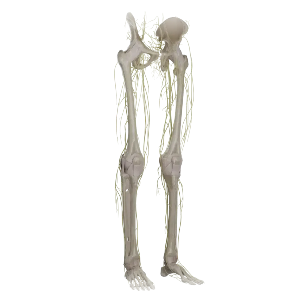
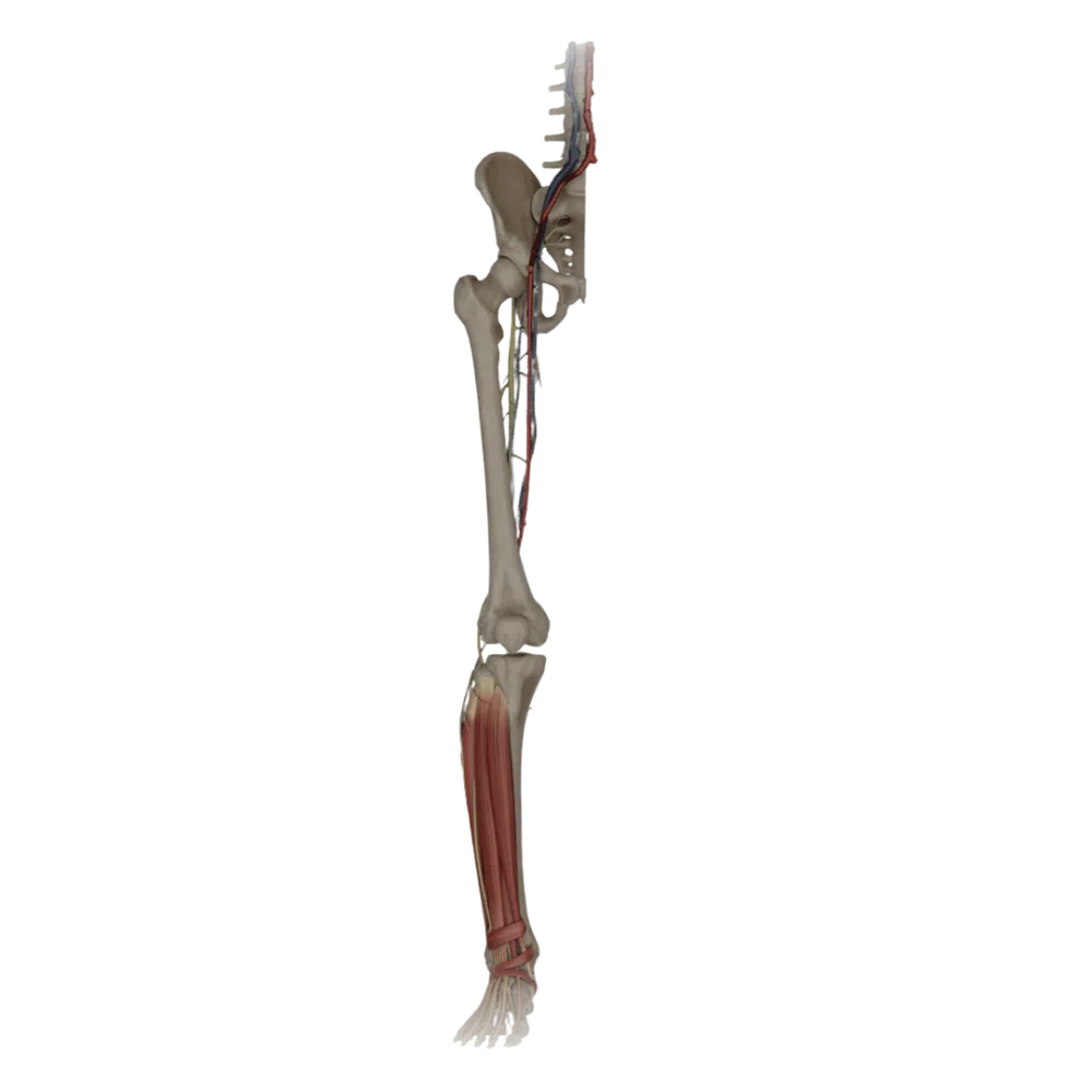
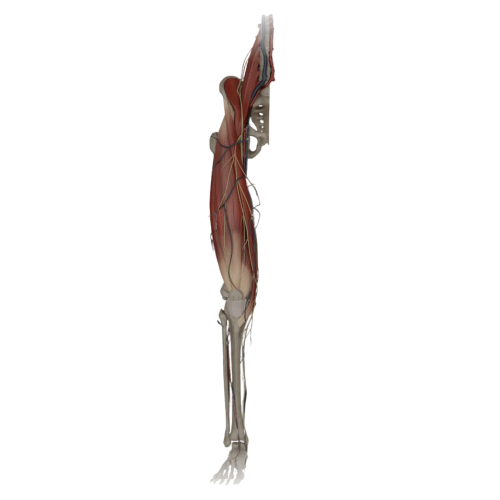
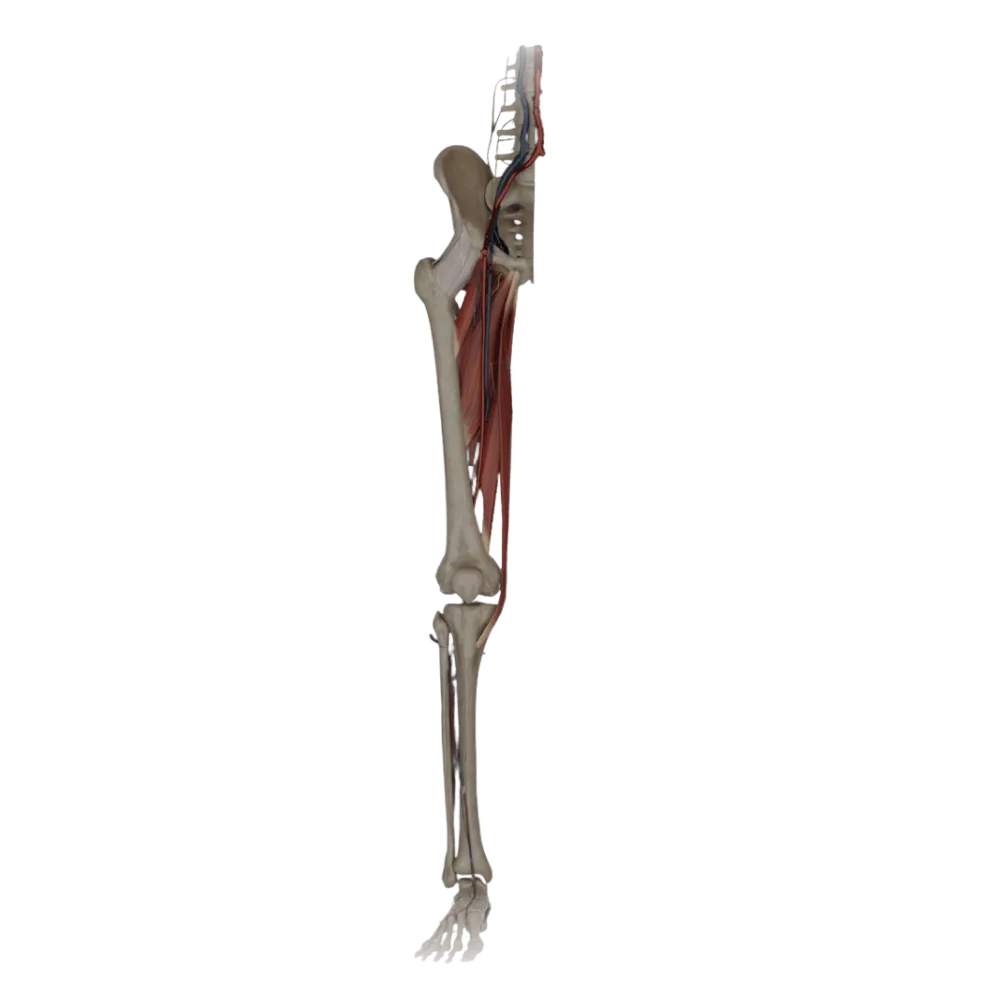

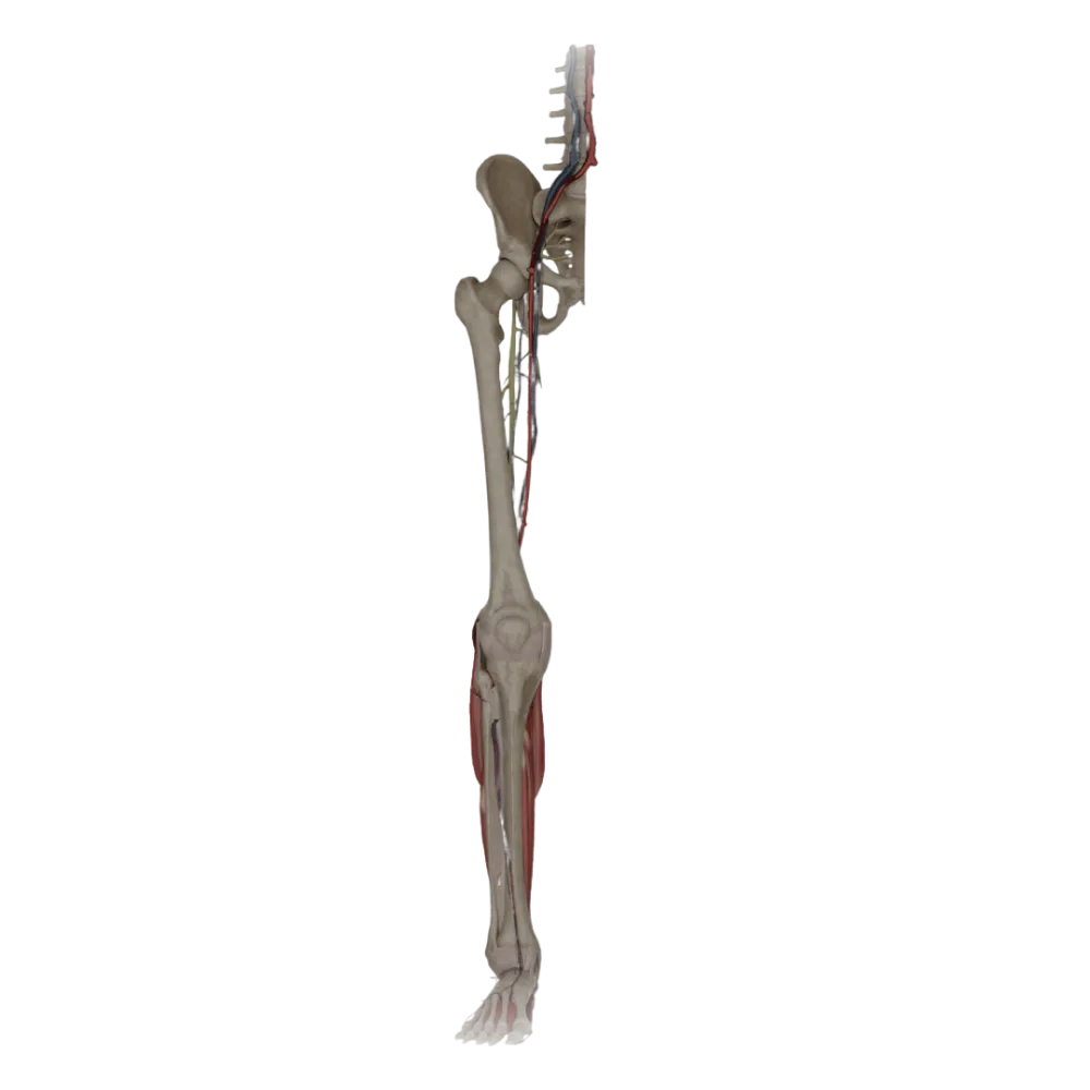
Anatomy test of the nerves of the lower limb.
Evaluate the knowledge of anatomy of the nerves of the lower limb. The test assesses their topography, course, branching, and specific zones of innervation of muscles and skin.
1. Which nerve exits onto the thigh through the muscular lacuna?
-
Obturator nerve.
The femoral nerve exits the pelvic cavity onto the thigh through the muscular lacuna along with the iliopsoas muscle.
-
Femoral nerve.
The femoral nerve exits the pelvic cavity onto the thigh through the muscular lacuna along with the iliopsoas muscle.
-
Ilioinguinal nerve
The femoral nerve exits the pelvic cavity onto the thigh through the muscular lacuna along with the iliopsoas muscle.
-
Sciatic nerve
The femoral nerve exits the pelvic cavity onto the thigh through the muscular lacuna along with the iliopsoas muscle.
-
I find it difficult to answer
The femoral nerve exits the pelvic cavity onto the thigh through the muscular lacuna along with the iliopsoas muscle.
2. Which muscle is NOT innervated by the obturator nerve?
-
Adductor brevis
The sartorius muscle is innervated by the femoral nerve, while the listed muscles belong to the medial group and are innervated by the obturator nerve.
-
Gracilis muscle.
The sartorius muscle is innervated by the femoral nerve, while the listed muscles belong to the medial group and are innervated by the obturator nerve.
-
Adductor longus
The sartorius muscle is innervated by the femoral nerve, while the listed muscles belong to the medial group and are innervated by the obturator nerve.
-
Sartorius muscle.
The sartorius muscle is innervated by the femoral nerve, while the listed muscles belong to the medial group and are innervated by the obturator nerve.
-
I find it difficult to answer
The sartorius muscle is innervated by the femoral nerve, while the listed muscles belong to the medial group and are innervated by the obturator nerve.
3. Through which foramen does the sciatic nerve leave the pelvic cavity?
-
Suprapiriform foramen
The sciatic nerve exits the pelvic cavity through the infrapiriform foramen, positioned laterally to the inferior gluteal vessels.
-
Infrapiriform foramen
The sciatic nerve exits the pelvic cavity through the infrapiriform foramen, positioned laterally to the inferior gluteal vessels.
-
Lesser sciatic foramen
The sciatic nerve exits the pelvic cavity through the infrapiriform foramen, positioned laterally to the inferior gluteal vessels.
-
Obturator canal.
The sciatic nerve exits the pelvic cavity through the infrapiriform foramen, positioned laterally to the inferior gluteal vessels.
-
I find it difficult to answer
The sciatic nerve exits the pelvic cavity through the infrapiriform foramen, positioned laterally to the inferior gluteal vessels.
4. What position does the tibial nerve occupy in the popliteal fossa relative to the popliteal vessels?
-
It lies most superficially and laterally.
In the popliteal fossa, the elements of the neurovascular bundle are arranged in surface depth order (lateral to medial): nerve, vein, artery.
-
It lies deeper than the artery and more medially.
In the popliteal fossa, the elements of the neurovascular bundle are arranged in surface depth order (lateral to medial): nerve, vein, artery.
-
It lies between the artery and the vein.
In the popliteal fossa, the elements of the neurovascular bundle are arranged in surface depth order (lateral to medial): nerve, vein, artery.
-
It lies the deepest and laterally.
In the popliteal fossa, the elements of the neurovascular bundle are arranged in surface depth order (lateral to medial): nerve, vein, artery.
-
I find it difficult to answer
In the popliteal fossa, the elements of the neurovascular bundle are arranged in surface depth order (lateral to medial): nerve, vein, artery.
5. Which nerve is the sural nerve (n. suralis) a branch of?
-
Only of the tibial nerve.
The sural nerve is formed in the lower third of the leg by the union of the medial sural cutaneous nerve and the lateral sural cutaneous nerve.
-
Only of the common fibular nerve.
The sural nerve is formed in the lower third of the leg by the union of the medial sural cutaneous nerve and the lateral sural cutaneous nerve.
-
Formed by the merger of branches of the tibial and common fibular nerves.
The sural nerve is formed in the lower third of the leg by the union of the medial sural cutaneous nerve and the lateral sural cutaneous nerve.
-
Saphenous nerve
The sural nerve is formed in the lower third of the leg by the union of the medial sural cutaneous nerve and the lateral sural cutaneous nerve.
-
I find it difficult to answer
The sural nerve is formed in the lower third of the leg by the union of the medial sural cutaneous nerve and the lateral sural cutaneous nerve.
6. Which muscles are innervated by the deep fibular nerve?
-
Muscles of the anterior compartment of the leg.
The deep fibular nerve descends alongside the interosseous membrane and innervates the anterior tibial muscle and long extensors of the toes.
-
Muscles of the lateral compartment of the leg.
The deep fibular nerve descends alongside the interosseous membrane and innervates the anterior tibial muscle and long extensors of the toes.
-
Muscles of the posterior compartment of the leg.
The deep fibular nerve descends alongside the interosseous membrane and innervates the anterior tibial muscle and long extensors of the toes.
-
Muscles of the plantar surface of the foot.
The deep fibular nerve descends alongside the interosseous membrane and innervates the anterior tibial muscle and long extensors of the toes.
-
I find it difficult to answer
The deep fibular nerve descends alongside the interosseous membrane and innervates the anterior tibial muscle and long extensors of the toes.
7. Which skin area is innervated by the superficial fibular nerve?
-
Skin of the sole
The superficial fibular nerve provides sensory innervation to most of the skin of the dorsum of the foot, except for the first interdigital space.
-
Skin of the medial side of the leg
The superficial fibular nerve provides sensory innervation to most of the skin of the dorsum of the foot, except for the first interdigital space.
-
Skin of the dorsum of the foot (except for the first interdigital space)
The superficial fibular nerve provides sensory innervation to most of the skin of the dorsum of the foot, except for the first interdigital space.
-
Skin of the posterior surface of the leg.
The superficial fibular nerve provides sensory innervation to most of the skin of the dorsum of the foot, except for the first interdigital space.
-
I find it difficult to answer
The superficial fibular nerve provides sensory innervation to most of the skin of the dorsum of the foot, except for the first interdigital space.
8. Through which structure does the saphenous nerve (n. saphenus) exit the adductor canal?
-
Upper opening of the canal
The saphenous nerve exits the adductor canal through its anterior wall (lamina vastoadductoria) along with the descending genicular artery.
-
Lower opening (tendinous hiatus)
The saphenous nerve exits the adductor canal through its anterior wall (lamina vastoadductoria) along with the descending genicular artery.
-
Obturator foramen
The saphenous nerve exits the adductor canal through its anterior wall (lamina vastoadductoria) along with the descending genicular artery.
-
Anterior opening (lamina vastoadductoria)
The saphenous nerve exits the adductor canal through its anterior wall (lamina vastoadductoria) along with the descending genicular artery.
-
I find it difficult to answer
The saphenous nerve exits the adductor canal through its anterior wall (lamina vastoadductoria) along with the descending genicular artery.
9. Which nerve accompanies the small saphenous vein on the posterior surface of the leg?
-
Saphenous nerve.
The sural nerve (n. suralis) descends along the posterior surface of the leg alongside the small saphenous vein, heading towards the lateral malleolus.
-
Sural nerve.
The sural nerve (n. suralis) descends along the posterior surface of the leg alongside the small saphenous vein, heading towards the lateral malleolus.
-
Lateral sural cutaneous nerve
The sural nerve (n. suralis) descends along the posterior surface of the leg alongside the small saphenous vein, heading towards the lateral malleolus.
-
Medial plantar nerve
The sural nerve (n. suralis) descends along the posterior surface of the leg alongside the small saphenous vein, heading towards the lateral malleolus.
-
I find it difficult to answer
The sural nerve (n. suralis) descends along the posterior surface of the leg alongside the small saphenous vein, heading towards the lateral malleolus.
10. How is the femoral nerve positioned in the femoral triangle relative to the femoral artery?
-
Medial to the artery
In the femoral triangle, the neurovascular bundle is organized from outside inwards: femoral nerve, femoral artery, femoral vein.
-
In front of the artery.
In the femoral triangle, the neurovascular bundle is organized from outside inwards: femoral nerve, femoral artery, femoral vein.
-
Lateral to the artery
In the femoral triangle, the neurovascular bundle is organized from outside inwards: femoral nerve, femoral artery, femoral vein.
-
Behind the artery
In the femoral triangle, the neurovascular bundle is organized from outside inwards: femoral nerve, femoral artery, femoral vein.
-
I find it difficult to answer
In the femoral triangle, the neurovascular bundle is organized from outside inwards: femoral nerve, femoral artery, femoral vein.
11. Which nerve innervates the gluteus maximus muscle?
-
Superior gluteal nerve
The inferior gluteal nerve exits through the infrapiriform foramen and branches within the thickness of the gluteus maximus muscle, providing its innervation.
-
Inferior gluteal nerve.
The inferior gluteal nerve exits through the infrapiriform foramen and branches within the thickness of the gluteus maximus muscle, providing its innervation.
-
Sciatic nerve
The inferior gluteal nerve exits through the infrapiriform foramen and branches within the thickness of the gluteus maximus muscle, providing its innervation.
-
Pudendal nerve
The inferior gluteal nerve exits through the infrapiriform foramen and branches within the thickness of the gluteus maximus muscle, providing its innervation.
-
I find it difficult to answer
The inferior gluteal nerve exits through the infrapiriform foramen and branches within the thickness of the gluteus maximus muscle, providing its innervation.
12. Which muscles does the medial plantar nerve innervate?
-
The muscle that abducts the hallux, the short flexor of the toes, and the first lumbrical.
The medial plantar nerve is functionally analogous to the median nerve in the hand, innervating the muscles of the thenar eminence and others.
-
Quadratus plantae
The medial plantar nerve is functionally analogous to the median nerve in the hand, innervating the muscles of the thenar eminence and others.
-
The muscle that abducts the little toe
The medial plantar nerve is functionally analogous to the median nerve in the hand, innervating the muscles of the thenar eminence and others.
-
All interosseous muscles
The medial plantar nerve is functionally analogous to the median nerve in the hand, innervating the muscles of the thenar eminence and others.
-
I find it difficult to answer
The medial plantar nerve is functionally analogous to the median nerve in the hand, innervating the muscles of the thenar eminence and others.
13. Which nerve wraps around the neck of the fibula, lying superficially and therefore susceptible to compression?
-
Tibial nerve.
The common fibular nerve wraps around the head and neck of the fibula externally, positioned directly beneath the fascia.
-
Deep fibular nerve.
The common fibular nerve wraps around the head and neck of the fibula externally, positioned directly beneath the fascia.
-
Superficial fibular nerve.
The common fibular nerve wraps around the head and neck of the fibula externally, positioned directly beneath the fascia.
-
Common fibular nerve.
The common fibular nerve wraps around the head and neck of the fibula externally, positioned directly beneath the fascia.
-
I find it difficult to answer
The common fibular nerve wraps around the head and neck of the fibula externally, positioned directly beneath the fascia.
14. Which plexus does the lateral femoral cutaneous nerve branch from?
-
Lumbar
The lateral femoral cutaneous nerve originates from the lumbar plexus (L2-L3) and heads toward the anterior superior iliac spine.
-
Cervical
The lateral femoral cutaneous nerve originates from the lumbar plexus (L2-L3) and heads toward the anterior superior iliac spine.
-
Brachial
The lateral femoral cutaneous nerve originates from the lumbar plexus (L2-L3) and heads toward the anterior superior iliac spine.
-
Sacral
The lateral femoral cutaneous nerve originates from the lumbar plexus (L2-L3) and heads toward the anterior superior iliac spine.
-
I find it difficult to answer
The lateral femoral cutaneous nerve originates from the lumbar plexus (L2-L3) and heads toward the anterior superior iliac spine.
15. What is the source of innervation for the gluteus medius and minimus muscles, as well as the tensor fasciae latae?
-
Superior gluteal nerve
The superior gluteal nerve exits the pelvis through the suprapiriform foramen and heads towards the gluteus medius and minimus muscles.
-
Inferior gluteal nerve.
The superior gluteal nerve exits the pelvis through the suprapiriform foramen and heads towards the gluteus medius and minimus muscles.
-
Femoral nerve.
The superior gluteal nerve exits the pelvis through the suprapiriform foramen and heads towards the gluteus medius and minimus muscles.
-
Obturator nerve.
The superior gluteal nerve exits the pelvis through the suprapiriform foramen and heads towards the gluteus medius and minimus muscles.
-
I find it difficult to answer
The superior gluteal nerve exits the pelvis through the suprapiriform foramen and heads towards the gluteus medius and minimus muscles.
16. Which nerve innervates the skin of the first interdigital space on the dorsum of the foot?
-
Sural nerve.
The deep fibular nerve, upon reaching the dorsum of the foot, divides into branches that innervate the adjacent surfaces of the first and second toes.
-
Deep fibular nerve.
The deep fibular nerve, upon reaching the dorsum of the foot, divides into branches that innervate the adjacent surfaces of the first and second toes.
-
Superficial fibular nerve.
The deep fibular nerve, upon reaching the dorsum of the foot, divides into branches that innervate the adjacent surfaces of the first and second toes.
-
Saphenous nerve.
The deep fibular nerve, upon reaching the dorsum of the foot, divides into branches that innervate the adjacent surfaces of the first and second toes.
-
I find it difficult to answer
The deep fibular nerve, upon reaching the dorsum of the foot, divides into branches that innervate the adjacent surfaces of the first and second toes.
17. How does the lateral femoral cutaneous nerve emerge onto the thigh?
-
Through the vascular lacuna
The lateral femoral cutaneous nerve passes under the inguinal ligament at a distance of 1-2 cm medial to the anterior superior iliac spine.
-
Through the muscular lacuna
The lateral femoral cutaneous nerve passes under the inguinal ligament at a distance of 1-2 cm medial to the anterior superior iliac spine.
-
Under the inguinal ligament, medial to the anterior superior iliac spine
The lateral femoral cutaneous nerve passes under the inguinal ligament at a distance of 1-2 cm medial to the anterior superior iliac spine.
-
Through the obturator canal
The lateral femoral cutaneous nerve passes under the inguinal ligament at a distance of 1-2 cm medial to the anterior superior iliac spine.
-
I find it difficult to answer
The lateral femoral cutaneous nerve passes under the inguinal ligament at a distance of 1-2 cm medial to the anterior superior iliac spine.
18. At what level does the sciatic nerve typically divide into its terminal branches (tibial and common fibular nerves)?
-
Level of the greater trochanter
In typical cases, the sciatic nerve divides into the tibial and common fibular nerves at the upper angle of the popliteal fossa.
-
Middle third of the thigh
In typical cases, the sciatic nerve divides into the tibial and common fibular nerves at the upper angle of the popliteal fossa.
-
Ankle joint
In typical cases, the sciatic nerve divides into the tibial and common fibular nerves at the upper angle of the popliteal fossa.
-
Upper angle of the popliteal fossa
In typical cases, the sciatic nerve divides into the tibial and common fibular nerves at the upper angle of the popliteal fossa.
-
I find it difficult to answer
In typical cases, the sciatic nerve divides into the tibial and common fibular nerves at the upper angle of the popliteal fossa.
19. Which nerve passes through the vascular lacuna?
-
Femoral nerve.
The femoral branch of the genitofemoral nerve travels onto the thigh through the vascular lacuna, positioned laterally and anterior to the artery.
-
Femoral branch of the genitofemoral nerve
The femoral branch of the genitofemoral nerve travels onto the thigh through the vascular lacuna, positioned laterally and anterior to the artery.
-
Obturator nerve.
The femoral branch of the genitofemoral nerve travels onto the thigh through the vascular lacuna, positioned laterally and anterior to the artery.
-
Ilioinguinal nerve
The femoral branch of the genitofemoral nerve travels onto the thigh through the vascular lacuna, positioned laterally and anterior to the artery.
-
I find it difficult to answer
The femoral branch of the genitofemoral nerve travels onto the thigh through the vascular lacuna, positioned laterally and anterior to the artery.
20. Which branch of the sacral plexus descends along the posterior surface of the thigh beneath the fascia lata, innervating the skin of this area?
-
Sciatic nerve
The posterior femoral cutaneous nerve exits through the infrapiriform foramen, descends, and innervates the skin of the posterior surface of the thigh.
-
Lateral femoral cutaneous nerve.
The posterior femoral cutaneous nerve exits through the infrapiriform foramen, descends, and innervates the skin of the posterior surface of the thigh.
-
Posterior femoral cutaneous nerve.
The posterior femoral cutaneous nerve exits through the infrapiriform foramen, descends, and innervates the skin of the posterior surface of the thigh.
-
Pudendal nerve
The posterior femoral cutaneous nerve exits through the infrapiriform foramen, descends, and innervates the skin of the posterior surface of the thigh.
-
I find it difficult to answer
The posterior femoral cutaneous nerve exits through the infrapiriform foramen, descends, and innervates the skin of the posterior surface of the thigh.
Retake this quiz?
Your current progress will be reset.
Nerves of the lower limb.
Tibial nerve.
0/20