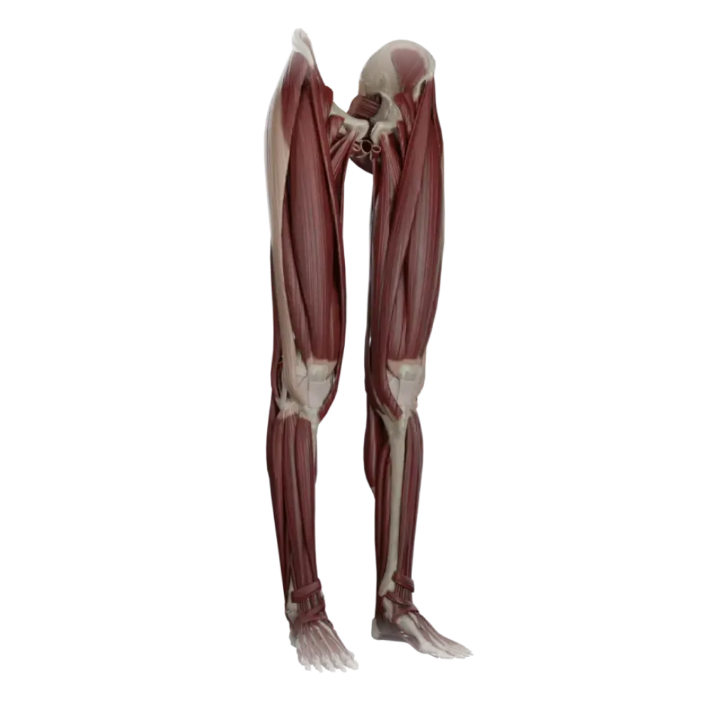

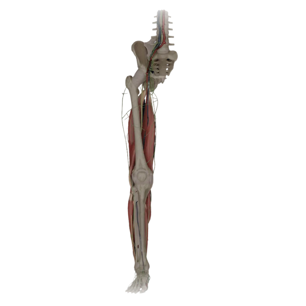
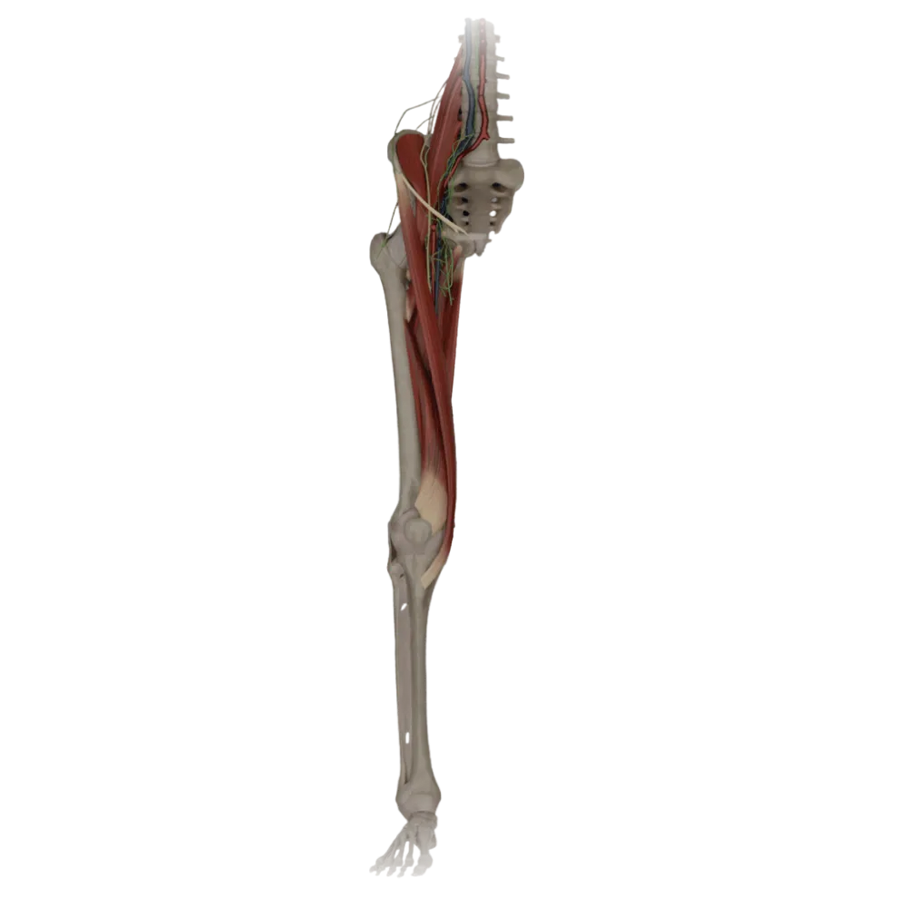
Thigh anatomy test
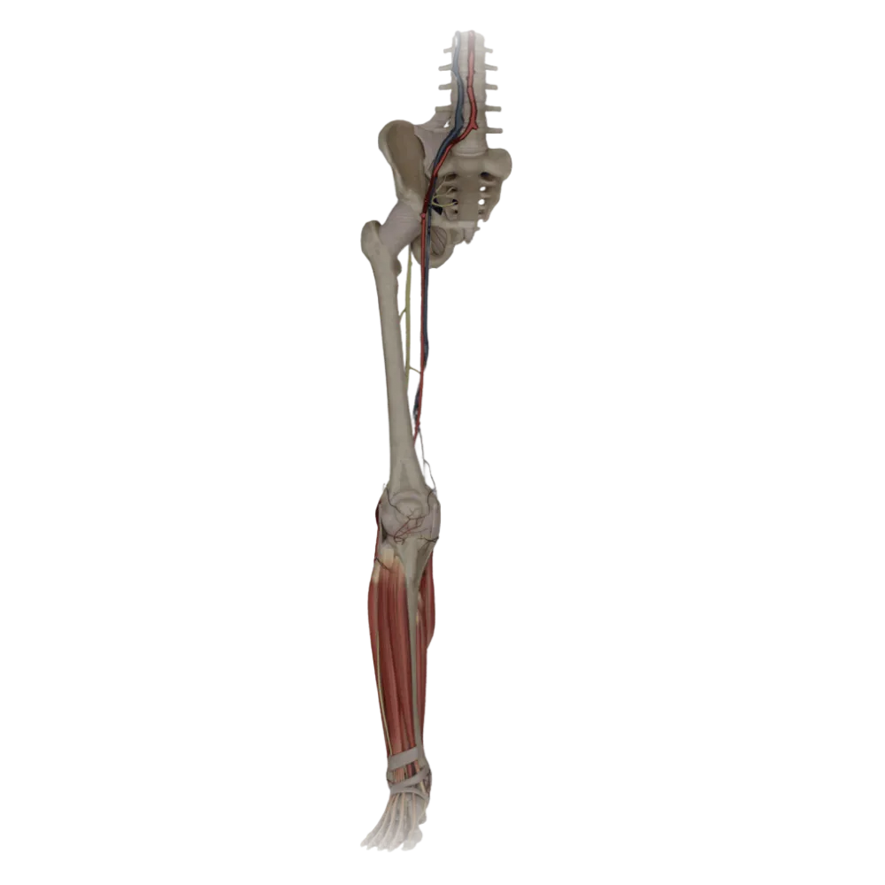
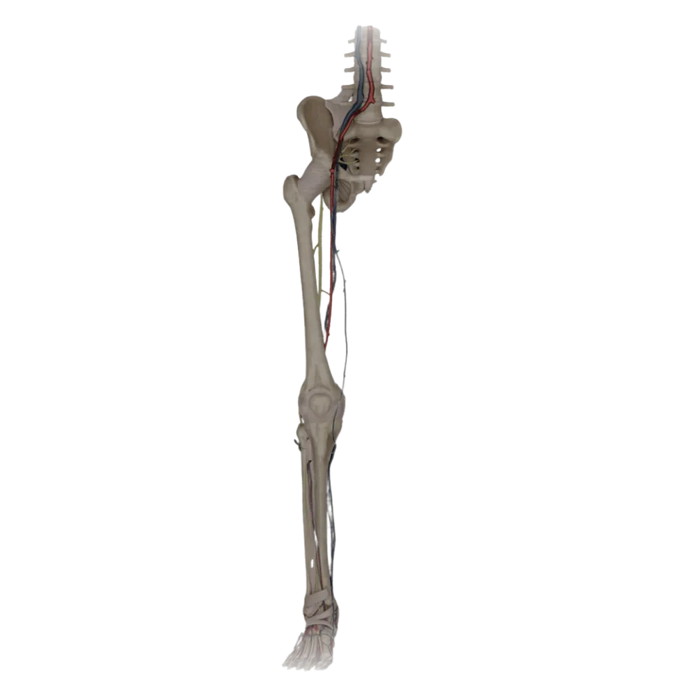
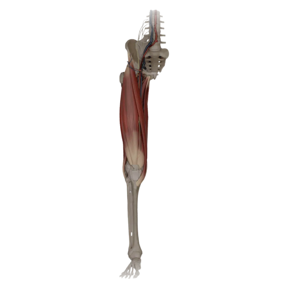
Evaluate your knowledge of thigh anatomy. The test assesses the boundaries and contents of the femoral triangle, muscular lacuna, and the adductor canal.
1/20
bold
text
1. Which structure forms the superior boundary of the femoral triangle?
-
Inguinal ligament
The femoral triangle (Scarpa's triangle) is bounded superiorly by the inguinal ligament.
-
Sartorius muscle.
The femoral triangle (Scarpa's triangle) is bounded superiorly by the inguinal ligament.
-
Adductor longus
The femoral triangle (Scarpa's triangle) is bounded superiorly by the inguinal ligament.
-
Pectineus muscle.
The femoral triangle (Scarpa's triangle) is bounded superiorly by the inguinal ligament.
-
I find it difficult to answer
The femoral triangle (Scarpa's triangle) is bounded superiorly by the inguinal ligament.
2. Which muscle forms the lateral boundary of the femoral triangle?
-
Rectus femoris.
The sartorius muscle's medial edge constitutes the femoral triangle's lateral boundary.
-
Sartorius muscle.
The sartorius muscle's medial edge constitutes the femoral triangle's lateral boundary.
-
Gracilis muscle.
The sartorius muscle's medial edge constitutes the femoral triangle's lateral boundary.
-
Tensor fasciae latae
The sartorius muscle's medial edge constitutes the femoral triangle's lateral boundary.
-
I find it difficult to answer
The sartorius muscle's medial edge constitutes the femoral triangle's lateral boundary.
3. What forms the medial boundary of the femoral triangle?
-
Gracilis muscle.
The medial boundary of the femoral triangle is formed by the medial edge of the adductor longus muscle.
-
Adductor magnus muscle.
The medial boundary of the femoral triangle is formed by the medial edge of the adductor longus muscle.
-
Adductor brevis
The medial boundary of the femoral triangle is formed by the medial edge of the adductor longus muscle.
-
Adductor longus
The medial boundary of the femoral triangle is formed by the medial edge of the adductor longus muscle.
-
I find it difficult to answer
The medial boundary of the femoral triangle is formed by the medial edge of the adductor longus muscle.
4. Which muscles form the floor of the femoral triangle (iliopubic fossa)?
-
Rectus and broad thigh muscles
The floor of the triangle, or iliopubic fossa, is formed by the iliopsoas laterally and the pectineus medially.
-
Iliopsoas and pectineus muscles
The floor of the triangle, or iliopubic fossa, is formed by the iliopsoas laterally and the pectineus medially.
-
External and internal obturator muscles
The floor of the triangle, or iliopubic fossa, is formed by the iliopsoas laterally and the pectineus medially.
-
Adductor magnus and longus muscles
The floor of the triangle, or iliopubic fossa, is formed by the iliopsoas laterally and the pectineus medially.
-
I find it difficult to answer
The floor of the triangle, or iliopubic fossa, is formed by the iliopsoas laterally and the pectineus medially.
5. In what sequence (from lateral to medial) are the major structures located in the vascular lacuna and the femoral triangle?
-
Vein, artery, nerve
Sequence of structures from lateral to medial: femoral nerve (in the muscular lacuna), femoral artery, femoral vein (in the vascular lacuna).
-
Nerve, vein, artery
Sequence of structures from lateral to medial: femoral nerve (in the muscular lacuna), femoral artery, femoral vein (in the vascular lacuna).
-
Nerve, artery, vein
Sequence of structures from lateral to medial: femoral nerve (in the muscular lacuna), femoral artery, femoral vein (in the vascular lacuna).
-
Artery, vein, nerve
Sequence of structures from lateral to medial: femoral nerve (in the muscular lacuna), femoral artery, femoral vein (in the vascular lacuna).
-
I find it difficult to answer
Sequence of structures from lateral to medial: femoral nerve (in the muscular lacuna), femoral artery, femoral vein (in the vascular lacuna).
6. Which structure forms the lateral wall of the adductor canal (Hunter's canal)?
-
Vastus medialis
The lateral wall of the adductor canal (m. vastus medialis) is formed by the vastus medialis.
-
Sartorius muscle.
The lateral wall of the adductor canal (m. vastus medialis) is formed by the vastus medialis.
-
Adductor longus
The lateral wall of the adductor canal (m. vastus medialis) is formed by the vastus medialis.
-
Biceps femoris muscle.
The lateral wall of the adductor canal (m. vastus medialis) is formed by the vastus medialis.
-
I find it difficult to answer
The lateral wall of the adductor canal (m. vastus medialis) is formed by the vastus medialis.
7. What forms the medial wall of the adductor canal?
-
Semitendinosus muscle.
The medial (posteromedial) wall is formed by the adductor magnus (m. adductor magnus).
-
Rectus femoris.
The medial (posteromedial) wall is formed by the adductor magnus (m. adductor magnus).
-
Pectineus muscle.
The medial (posteromedial) wall is formed by the adductor magnus (m. adductor magnus).
-
Adductor magnus muscle.
The medial (posteromedial) wall is formed by the adductor magnus (m. adductor magnus).
-
I find it difficult to answer
The medial (posteromedial) wall is formed by the adductor magnus (m. adductor magnus).
8. Which structure forms the anterior wall of the adductor canal?
-
Fascia lata
The anterior wall is the fibrous plate stretched between the vastus medialis and adductor magnus muscles.
-
Sartorius muscle.
The anterior wall is the fibrous plate stretched between the vastus medialis and adductor magnus muscles.
-
Fibrous plate (lamina vastoadductoria)
The anterior wall is the fibrous plate stretched between the vastus medialis and adductor magnus muscles.
-
Iliotibial tract
The anterior wall is the fibrous plate stretched between the vastus medialis and adductor magnus muscles.
-
I find it difficult to answer
The anterior wall is the fibrous plate stretched between the vastus medialis and adductor magnus muscles.
9. Which elements pass through the entire length of the adductor canal?
-
Femoral artery and obturator nerve
The femoral artery and femoral vein pass through the adductor canal heading to the popliteal fossa.
-
Femoral artery and femoral vein
The femoral artery and femoral vein pass through the adductor canal heading to the popliteal fossa.
-
Deep femoral artery and sciatic nerve
The femoral artery and femoral vein pass through the adductor canal heading to the popliteal fossa.
-
Great saphenous vein and saphenous nerve
The femoral artery and femoral vein pass through the adductor canal heading to the popliteal fossa.
-
I find it difficult to answer
The femoral artery and femoral vein pass through the adductor canal heading to the popliteal fossa.
10. Which nerve enters the adductor canal but exits through its anterior opening without reaching the popliteal fossa?
-
Saphenous nerve (n. saphenus)
The saphenous nerve pierces the anterior wall of the canal (lamina vastoadductoria) and emerges on the leg.
-
Obturator nerve.
The saphenous nerve pierces the anterior wall of the canal (lamina vastoadductoria) and emerges on the leg.
-
Femoral nerve.
The saphenous nerve pierces the anterior wall of the canal (lamina vastoadductoria) and emerges on the leg.
-
Sciatic nerve
The saphenous nerve pierces the anterior wall of the canal (lamina vastoadductoria) and emerges on the leg.
-
I find it difficult to answer
The saphenous nerve pierces the anterior wall of the canal (lamina vastoadductoria) and emerges on the leg.
11. Which artery exits the adductor canal alongside the saphenous nerve through its anterior opening?
-
Lateral circumflex femoral artery
The descending genicular artery arises from the femoral artery in the canal and exits through the anterior wall.
-
Medial circumflex femoral artery
The descending genicular artery arises from the femoral artery in the canal and exits through the anterior wall.
-
Descending genicular artery
The descending genicular artery arises from the femoral artery in the canal and exits through the anterior wall.
-
Perforating artery
The descending genicular artery arises from the femoral artery in the canal and exits through the anterior wall.
-
I find it difficult to answer
The descending genicular artery arises from the femoral artery in the canal and exits through the anterior wall.
12. Where does the lower opening of the adductor canal (hiatus adductorius) lead?
-
On the anterior surface of the shin
The hiatus adductorius of the adductor magnus muscle (hiatus adductorius) serves as the canal’s exit leading to the popliteal fossa.
-
Into the obturator canal
The hiatus adductorius of the adductor magnus muscle (hiatus adductorius) serves as the canal’s exit leading to the popliteal fossa.
-
Into the gluteal region
The hiatus adductorius of the adductor magnus muscle (hiatus adductorius) serves as the canal’s exit leading to the popliteal fossa.
-
Into the popliteal fossa
The hiatus adductorius of the adductor magnus muscle (hiatus adductorius) serves as the canal’s exit leading to the popliteal fossa.
-
I find it difficult to answer
The hiatus adductorius of the adductor magnus muscle (hiatus adductorius) serves as the canal’s exit leading to the popliteal fossa.
13. Which ligament divides the space under the inguinal ligament into the muscular and vascular lacunae?
-
Lacunar ligament.
The iliopectineal arch extends between the inguinal ligament and the iliopectineal eminence, dividing the lacunae.
-
Iliopectineal arch
The iliopectineal arch extends between the inguinal ligament and the iliopectineal eminence, dividing the lacunae.
-
Pectineal ligament.
The iliopectineal arch extends between the inguinal ligament and the iliopectineal eminence, dividing the lacunae.
-
Sacrotuberous ligament
The iliopectineal arch extends between the inguinal ligament and the iliopectineal eminence, dividing the lacunae.
-
I find it difficult to answer
The iliopectineal arch extends between the inguinal ligament and the iliopectineal eminence, dividing the lacunae.
14. What passes through the muscular lacuna?
-
Femoral artery and vein
The iliopsoas muscle and femoral nerve exit to the thigh through the muscular lacuna.
-
Pectineus muscle and obturator nerve
The iliopsoas muscle and femoral nerve exit to the thigh through the muscular lacuna.
-
Iliopsoas muscle and femoral nerve
The iliopsoas muscle and femoral nerve exit to the thigh through the muscular lacuna.
-
Sartorius muscle and lateral cutaneous nerve of the thigh
The iliopsoas muscle and femoral nerve exit to the thigh through the muscular lacuna.
-
I find it difficult to answer
The iliopsoas muscle and femoral nerve exit to the thigh through the muscular lacuna.
15. What forms the medial wall of the deep femoral ring?
-
Lacunar ligament.
The lacunar ligament forms the medial boundary of the deep femoral ring (anulus femoralis profundus).
-
Pectineal ligament.
The lacunar ligament forms the medial boundary of the deep femoral ring (anulus femoralis profundus).
-
Inguinal ligament
The lacunar ligament forms the medial boundary of the deep femoral ring (anulus femoralis profundus).
-
Femoral vein
The lacunar ligament forms the medial boundary of the deep femoral ring (anulus femoralis profundus).
-
I find it difficult to answer
The lacunar ligament forms the medial boundary of the deep femoral ring (anulus femoralis profundus).
16. What bounds the deep femoral ring laterally?
-
Femoral artery.
The lateral boundary of the femoral ring is the wall of the femoral vein.
-
Femoral vein
The lateral boundary of the femoral ring is the wall of the femoral vein.
-
Femoral nerve.
The lateral boundary of the femoral ring is the wall of the femoral vein.
-
Iliopectineal arch
The lateral boundary of the femoral ring is the wall of the femoral vein.
-
I find it difficult to answer
The lateral boundary of the femoral ring is the wall of the femoral vein.
17. What forms the posterior wall of the deep femoral ring?
-
Inguinal ligament
The posterior boundary of the ring is formed by the pectineal ligament located on the pubic crest.
-
Transversalis fascia
The posterior boundary of the ring is formed by the pectineal ligament located on the pubic crest.
-
Lacunar ligament.
The posterior boundary of the ring is formed by the pectineal ligament located on the pubic crest.
-
Pectineal ligament (also known as the ligament of Cooper)
The posterior boundary of the ring is formed by the pectineal ligament located on the pubic crest.
-
I find it difficult to answer
The posterior boundary of the ring is formed by the pectineal ligament located on the pubic crest.
18. What is typically located within the deep femoral ring?
-
Deep inguinal lymph node (also known as the node of Pirogov-Rosenmuller)
The deep femoral ring is filled with loose connective tissue and a prominent lymph node of Pirogov-Rosenmuller.
-
Femoral nerve.
The deep femoral ring is filled with loose connective tissue and a prominent lymph node of Pirogov-Rosenmuller.
-
Femoral artery.
The deep femoral ring is filled with loose connective tissue and a prominent lymph node of Pirogov-Rosenmuller.
-
Great saphenous vein
The deep femoral ring is filled with loose connective tissue and a prominent lymph node of Pirogov-Rosenmuller.
-
I find it difficult to answer
The deep femoral ring is filled with loose connective tissue and a prominent lymph node of Pirogov-Rosenmuller.
19. What is the name of the superficial opening of the femoral canal in the fascia lata of the thigh?
-
Superficial inguinal ring
The superficial ring of the femoral canal is the saphenous opening (hiatus saphenus), the edge of which is crescent-shaped.
-
Saphenous opening (hiatus saphenus)
The superficial ring of the femoral canal is the saphenous opening (hiatus saphenus), the edge of which is crescent-shaped.
-
Obturator canal.
The superficial ring of the femoral canal is the saphenous opening (hiatus saphenus), the edge of which is crescent-shaped.
-
Vascular lacuna.
The superficial ring of the femoral canal is the saphenous opening (hiatus saphenus), the edge of which is crescent-shaped.
-
I find it difficult to answer
The superficial ring of the femoral canal is the saphenous opening (hiatus saphenus), the edge of which is crescent-shaped.
20. Which vein penetrates the cribiform fascia at the region of the saphenous opening, draining into the femoral vein?
-
Small saphenous vein
The great saphenous vein of the leg passes through the saphenous opening and drains into the femoral vein.
-
External iliac vein
The great saphenous vein of the leg passes through the saphenous opening and drains into the femoral vein.
-
Great saphenous vein
The great saphenous vein of the leg passes through the saphenous opening and drains into the femoral vein.
-
Obturator veins
The great saphenous vein of the leg passes through the saphenous opening and drains into the femoral vein.
-
I find it difficult to answer
The great saphenous vein of the leg passes through the saphenous opening and drains into the femoral vein.
Retake this quiz?
Your current progress will be reset.