
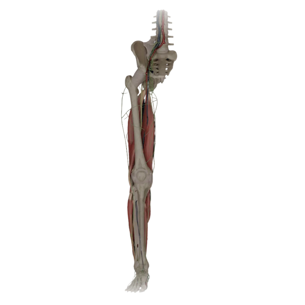
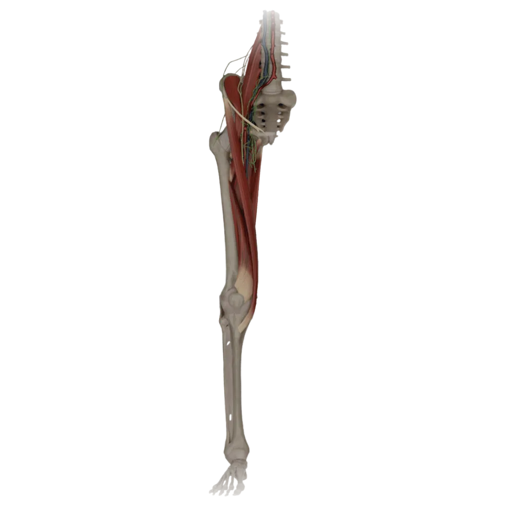
Anatomy test of the leg and foot
Evaluate the knowledge of leg and foot topography. The test examines the popliteal fossa, cruro-popliteal canal, musculoperoneal canals, and the sole.
1/20
bold
text
1. Which muscle borders the popliteal fossa superiorly and laterally?
-
Semimembranosus muscle.
The popliteal fossa is bordered superiorly and laterally by the biceps femoris, and superiorly and medially by the semimembranosus and semitendinosus muscles.
-
Biceps femoris muscle.
The popliteal fossa is bordered superiorly and laterally by the biceps femoris, and superiorly and medially by the semimembranosus and semitendinosus muscles.
-
Sartorius muscle.
The popliteal fossa is bordered superiorly and laterally by the biceps femoris, and superiorly and medially by the semimembranosus and semitendinosus muscles.
-
Gastrocnemius muscle.
The popliteal fossa is bordered superiorly and laterally by the biceps femoris, and superiorly and medially by the semimembranosus and semitendinosus muscles.
-
I find it difficult to answer
The popliteal fossa is bordered superiorly and laterally by the biceps femoris, and superiorly and medially by the semimembranosus and semitendinosus muscles.
2. What forms the floor of the popliteal fossa?
-
Long head of biceps femoris
The floor of the popliteal fossa (fossa poplitea) is formed by the popliteal surface of the femur and the posterior wall of the knee joint capsule
-
Fascia of the leg
The floor of the popliteal fossa (fossa poplitea) is formed by the popliteal surface of the femur and the posterior wall of the knee joint capsule
-
Soleus muscle.
The floor of the popliteal fossa (fossa poplitea) is formed by the popliteal surface of the femur and the posterior wall of the knee joint capsule
-
Popliteal surface of the femoral bone and knee joint capsule
The floor of the popliteal fossa (fossa poplitea) is formed by the popliteal surface of the femur and the posterior wall of the knee joint capsule
-
I find it difficult to answer
The floor of the popliteal fossa (fossa poplitea) is formed by the popliteal surface of the femur and the posterior wall of the knee joint capsule
3. What is the syntopic relationship of the vascular-nervous bundle elements in the popliteal fossa (from superficial to deep and medial)?
-
Tibial nerve, popliteal vein, popliteal artery
According to the NEVA rule (from superficial to deep and medially): Nerve (tibial), Vein (popliteal), Artery (popliteal).
-
Popliteal artery, popliteal vein, tibial nerve
According to the NEVA rule (from superficial to deep and medially): Nerve (tibial), Vein (popliteal), Artery (popliteal).
-
Popliteal vein, tibial nerve, popliteal artery
According to the NEVA rule (from superficial to deep and medially): Nerve (tibial), Vein (popliteal), Artery (popliteal).
-
Common fibular nerve, popliteal vein, popliteal artery
According to the NEVA rule (from superficial to deep and medially): Nerve (tibial), Vein (popliteal), Artery (popliteal).
-
I find it difficult to answer
According to the NEVA rule (from superficial to deep and medially): Nerve (tibial), Vein (popliteal), Artery (popliteal).
4. What forms the anterior wall of the cruro-popliteal canal (Gruber's canal)?
-
Soleus muscle
The anterior wall of the canalis cruropopliteus is formed by the tibialis posterior muscle, and the posterior by the soleus muscle.
-
Flexor digitorum longus
The anterior wall of the canalis cruropopliteus is formed by the tibialis posterior muscle, and the posterior by the soleus muscle.
-
Tibialis posterior muscle
The anterior wall of the canalis cruropopliteus is formed by the tibialis posterior muscle, and the posterior by the soleus muscle.
-
Tibialis anterior muscle
The anterior wall of the canalis cruropopliteus is formed by the tibialis posterior muscle, and the posterior by the soleus muscle.
-
I find it difficult to answer
The anterior wall of the canalis cruropopliteus is formed by the tibialis posterior muscle, and the posterior by the soleus muscle.
5. What limits the upper opening of the cruro-popliteal canal?
-
Medial and lateral heads of the gastrocnemius muscle
The upper (entry) opening of Gruber's canal is limited anteriorly by the m. popliteus, and posteriorly by the arcus tendineus m. solei
-
Popliteus muscle anteriorly and tendinous arch of the soleus muscle posteriorly
The upper (entry) opening of Gruber's canal is limited anteriorly by the m. popliteus, and posteriorly by the arcus tendineus m. solei
-
Interosseous membrane and tibialis posterior muscle
The upper (entry) opening of Gruber's canal is limited anteriorly by the m. popliteus, and posteriorly by the arcus tendineus m. solei
-
Flexor hallucis longus and fibula
The upper (entry) opening of Gruber's canal is limited anteriorly by the m. popliteus, and posteriorly by the arcus tendineus m. solei
-
I find it difficult to answer
The upper (entry) opening of Gruber's canal is limited anteriorly by the m. popliteus, and posteriorly by the arcus tendineus m. solei
6. What structure passes through the anterior opening of the cruro-popliteal canal in the interosseous membrane?
-
Posterior tibial artery.
Through the anterior opening at the top of the interosseous membrane the a. emerges on the anterior surface of the leg. Tibialis anterior.
-
Deep fibular nerve.
Through the anterior opening at the top of the interosseous membrane the a. emerges on the anterior surface of the leg. Tibialis anterior.
-
Superficial fibular nerve.
Through the anterior opening at the top of the interosseous membrane the a. emerges on the anterior surface of the leg. Tibialis anterior.
-
Anterior tibial artery.
Through the anterior opening at the top of the interosseous membrane the a. emerges on the anterior surface of the leg. Tibialis anterior.
-
I find it difficult to answer
Through the anterior opening at the top of the interosseous membrane the a. emerges on the anterior surface of the leg. Tibialis anterior.
7. What nerve passes in the upper musculoperoneal canal?
-
Superficial fibular nerve.
In the canalis musculoperoneus superior passes the superficial fibular nerve, which then pierces the fascia of the leg.
-
Deep fibular nerve.
In the canalis musculoperoneus superior passes the superficial fibular nerve, which then pierces the fascia of the leg.
-
Sural nerve.
In the canalis musculoperoneus superior passes the superficial fibular nerve, which then pierces the fascia of the leg.
-
Tibial nerve.
In the canalis musculoperoneus superior passes the superficial fibular nerve, which then pierces the fascia of the leg.
-
I find it difficult to answer
In the canalis musculoperoneus superior passes the superficial fibular nerve, which then pierces the fascia of the leg.
8. What structures form the walls of the lower musculoperoneal canal?
-
Tibia and flexor digitorum longus
The lower musculoperoneal canal is located in the lower third of the leg between the fibula and m. flexor hallucis longus
-
Fibula and fibularis longus muscle
The lower musculoperoneal canal is located in the lower third of the leg between the fibula and m. flexor hallucis longus
-
Fibula (laterally) and flexor hallucis longus (medially)
The lower musculoperoneal canal is located in the lower third of the leg between the fibula and m. flexor hallucis longus
-
Interosseous membrane and tibialis posterior muscle
The lower musculoperoneal canal is located in the lower third of the leg between the fibula and m. flexor hallucis longus
-
I find it difficult to answer
The lower musculoperoneal canal is located in the lower third of the leg between the fibula and m. flexor hallucis longus
9. What is the content of the lower musculoperoneal canal?
-
Anterior tibial artery.
In the lower musculoperoneal canal (a branch of the cruro-popliteal), fibular vessels pass (a. et vv. fibulares).
-
Fibular artery and accompanying veins
In the lower musculoperoneal canal (a branch of the cruro-popliteal), fibular vessels pass (a. et vv. fibulares).
-
Common fibular nerve.
In the lower musculoperoneal canal (a branch of the cruro-popliteal), fibular vessels pass (a. et vv. fibulares).
-
Saphenous nerve.
In the lower musculoperoneal canal (a branch of the cruro-popliteal), fibular vessels pass (a. et vv. fibulares).
-
I find it difficult to answer
In the lower musculoperoneal canal (a branch of the cruro-popliteal), fibular vessels pass (a. et vv. fibulares).
10. Where is the medial plantar groove (sulcus plantaris medialis) located?
-
Between flexor digitorum brevis and abductor digiti minimi muscle
The medial plantar groove is located between m. abductor hallucis medially and m. flexor digitorum brevis laterally.
-
On the dorsum of the foot between the tendons of extensors
The medial plantar groove is located between m. abductor hallucis medially and m. flexor digitorum brevis laterally.
-
Between the medial malleolus and the calcaneus
The medial plantar groove is located between m. abductor hallucis medially and m. flexor digitorum brevis laterally.
-
Between muscle that abducts the great toe and flexor digitorum brevis
The medial plantar groove is located between m. abductor hallucis medially and m. flexor digitorum brevis laterally.
-
I find it difficult to answer
The medial plantar groove is located between m. abductor hallucis medially and m. flexor digitorum brevis laterally.
11. What formation passes in the medial plantar groove?
-
Medial plantar vascular-nervous bundle (artery, veins and nerve)
In the sulcus plantaris medialis are located a., vv., et n. plantares mediales, which supply blood and innervate the medial part of the sole.
-
Lateral plantar artery
In the sulcus plantaris medialis are located a., vv., et n. plantares mediales, which supply blood and innervate the medial part of the sole.
-
The tendon of the fibularis longus muscle
In the sulcus plantaris medialis are located a., vv., et n. plantares mediales, which supply blood and innervate the medial part of the sole.
-
The deep plantar arch
In the sulcus plantaris medialis are located a., vv., et n. plantares mediales, which supply blood and innervate the medial part of the sole.
-
I find it difficult to answer
In the sulcus plantaris medialis are located a., vv., et n. plantares mediales, which supply blood and innervate the medial part of the sole.
12. What muscles delimit the lateral plantar groove?
-
Flexor digitorum longus and quadratus plantae muscle
The lateral plantar groove is delimited by m. flexor digitorum brevis and m. abductor digiti minimi
-
Flexor hallucis brevis and adductor hallucis
The lateral plantar groove is delimited by m. flexor digitorum brevis and m. abductor digiti minimi
-
Flexor digitorum brevis (medially) and abductor digiti minimi of the foot (laterally)
The lateral plantar groove is delimited by m. flexor digitorum brevis and m. abductor digiti minimi
-
Interosseous muscles and lumbrical muscles
The lateral plantar groove is delimited by m. flexor digitorum brevis and m. abductor digiti minimi
-
I find it difficult to answer
The lateral plantar groove is delimited by m. flexor digitorum brevis and m. abductor digiti minimi
13. Which artery is located in the lateral plantar groove?
-
Medial plantar artery
In the lateral groove passes a. plantaris lateralis, accompanied by the homonymous veins and nerve.
-
Lateral plantar artery
In the lateral groove passes a. plantaris lateralis, accompanied by the homonymous veins and nerve.
-
Dorsalis pedis artery
In the lateral groove passes a. plantaris lateralis, accompanied by the homonymous veins and nerve.
-
Perforating branch of the fibular artery
In the lateral groove passes a. plantaris lateralis, accompanied by the homonymous veins and nerve.
-
I find it difficult to answer
In the lateral groove passes a. plantaris lateralis, accompanied by the homonymous veins and nerve.
14. Where is the flexor retinaculum located, under which the tendons of the leg muscles pass?
-
On the lateral surface of the ankle joint
The flexor retinaculum stretches from the medial malleolus to the calcaneus, forming the malleolar canal.
-
On the anterior surface of the ankle joint
The flexor retinaculum stretches from the medial malleolus to the calcaneus, forming the malleolar canal.
-
In the popliteal fossa
The flexor retinaculum stretches from the medial malleolus to the calcaneus, forming the malleolar canal.
-
Between the medial malleolus and the calcaneus
The flexor retinaculum stretches from the medial malleolus to the calcaneus, forming the malleolar canal.
-
I find it difficult to answer
The flexor retinaculum stretches from the medial malleolus to the calcaneus, forming the malleolar canal.
15. What is the order of the deep leg muscle tendons posterior to the medial malleolus (from anterior to posterior)?
-
Tibialis posterior muscle, flexor digitorum longus, flexor hallucis longus
Topographic order from anterior to posterior under the flexor retinaculum: m. tibialis posterior, m. flexor digitorum longus, m. flexor hallucis longus
-
Flexor digitorum longus, flexor hallucis longus, tibialis posterior muscle
Topographic order from anterior to posterior under the flexor retinaculum: m. tibialis posterior, m. flexor digitorum longus, m. flexor hallucis longus
-
Flexor hallucis longus, tibialis posterior muscle, flexor digitorum longus
Topographic order from anterior to posterior under the flexor retinaculum: m. tibialis posterior, m. flexor digitorum longus, m. flexor hallucis longus
-
Tibialis anterior muscle, extensor digitorum longus, extensor hallucis longus
Topographic order from anterior to posterior under the flexor retinaculum: m. tibialis posterior, m. flexor digitorum longus, m. flexor hallucis longus
-
I find it difficult to answer
Topographic order from anterior to posterior under the flexor retinaculum: m. tibialis posterior, m. flexor digitorum longus, m. flexor hallucis longus
16. Which structure, exiting the popliteal fossa, lies in the groove between the heads of the gastrocnemius muscle along with the small saphenous vein?
-
Saphenous nerve.
The medial sural cutaneous nerve (n. cutaneus surae medialis) descends on the leg accompanied by v. saphena parva
-
Deep fibular nerve.
The medial sural cutaneous nerve (n. cutaneus surae medialis) descends on the leg accompanied by v. saphena parva
-
The medial sural cutaneous nerve (a branch of the tibial nerve)
The medial sural cutaneous nerve (n. cutaneus surae medialis) descends on the leg accompanied by v. saphena parva
-
Lateral plantar nerve
The medial sural cutaneous nerve (n. cutaneus surae medialis) descends on the leg accompanied by v. saphena parva
-
I find it difficult to answer
The medial sural cutaneous nerve (n. cutaneus surae medialis) descends on the leg accompanied by v. saphena parva
17. Which structure forms the posterior wall of the cruro-popliteal canal?
-
Flexor pollicis longus
The posterior wall of Gruber's canal is served by the anterior surface of the soleus muscle (m. soleus).
-
Soleus muscle.
The posterior wall of Gruber's canal is served by the anterior surface of the soleus muscle (m. soleus).
-
Gastrocnemius muscle.
The posterior wall of Gruber's canal is served by the anterior surface of the soleus muscle (m. soleus).
-
Popliteal muscle.
The posterior wall of Gruber's canal is served by the anterior surface of the soleus muscle (m. soleus).
-
I find it difficult to answer
The posterior wall of Gruber's canal is served by the anterior surface of the soleus muscle (m. soleus).
18. What emerges through the lower opening of the cruro-popliteal canal (between the Achilles tendon and the medial malleolus)?
-
Anterior tibial artery and deep fibular nerve
Through the lower opening of the canal onto the sole (into the malleolar canal) exit a. et vv. tibiales posteriores and n. tibialis
-
Fibular artery and superficial fibular nerve
Through the lower opening of the canal onto the sole (into the malleolar canal) exit a. et vv. tibiales posteriores and n. tibialis
-
Popliteal vein and common fibular nerve
Through the lower opening of the canal onto the sole (into the malleolar canal) exit a. et vv. tibiales posteriores and n. tibialis
-
Posterior tibial artery, accompanying veins, and tibial nerve
Through the lower opening of the canal onto the sole (into the malleolar canal) exit a. et vv. tibiales posteriores and n. tibialis
-
I find it difficult to answer
Through the lower opening of the canal onto the sole (into the malleolar canal) exit a. et vv. tibiales posteriores and n. tibialis
19. In the formation of the plantar arterial arch (arcus plantaris), the key role is played by:
-
Lateral plantar artery
The plantar arch is formed by the anastomosis of a large artery. Plantaris lateralis with deep plantar branch of dorsalis pedis artery
-
Medial plantar artery
The plantar arch is formed by the anastomosis of a large artery. Plantaris lateralis with deep plantar branch of dorsalis pedis artery
-
Perforating branch of the fibular artery
The plantar arch is formed by the anastomosis of a large artery. Plantaris lateralis with deep plantar branch of dorsalis pedis artery
-
Lateral tarsal artery
The plantar arch is formed by the anastomosis of a large artery. Plantaris lateralis with deep plantar branch of dorsalis pedis artery
-
I find it difficult to answer
The plantar arch is formed by the anastomosis of a large artery. Plantaris lateralis with deep plantar branch of dorsalis pedis artery
20. Which neurovascular bundle passes on the dorsum of the foot under the inferior extensor retinaculum?
-
Medial plantar artery and tibial nerve
On the dorsum of the foot under the retinaculum mm. extensorum inferius. Topographically pass Dorsalis pedis and n. Fibularis profundus
-
Superficial fibular nerve and small saphenous vein
On the dorsum of the foot under the retinaculum mm. extensorum inferius. Topographically pass Dorsalis pedis and n. Fibularis profundus
-
Dorsalis pedis artery and deep fibular nerve
On the dorsum of the foot under the retinaculum mm. extensorum inferius. Topographically pass Dorsalis pedis and n. Fibularis profundus
-
Sural nerve and lateral malleolar artery
On the dorsum of the foot under the retinaculum mm. extensorum inferius. Topographically pass Dorsalis pedis and n. Fibularis profundus
-
I find it difficult to answer
On the dorsum of the foot under the retinaculum mm. extensorum inferius. Topographically pass Dorsalis pedis and n. Fibularis profundus
Retake this quiz?
Your current progress will be reset.