
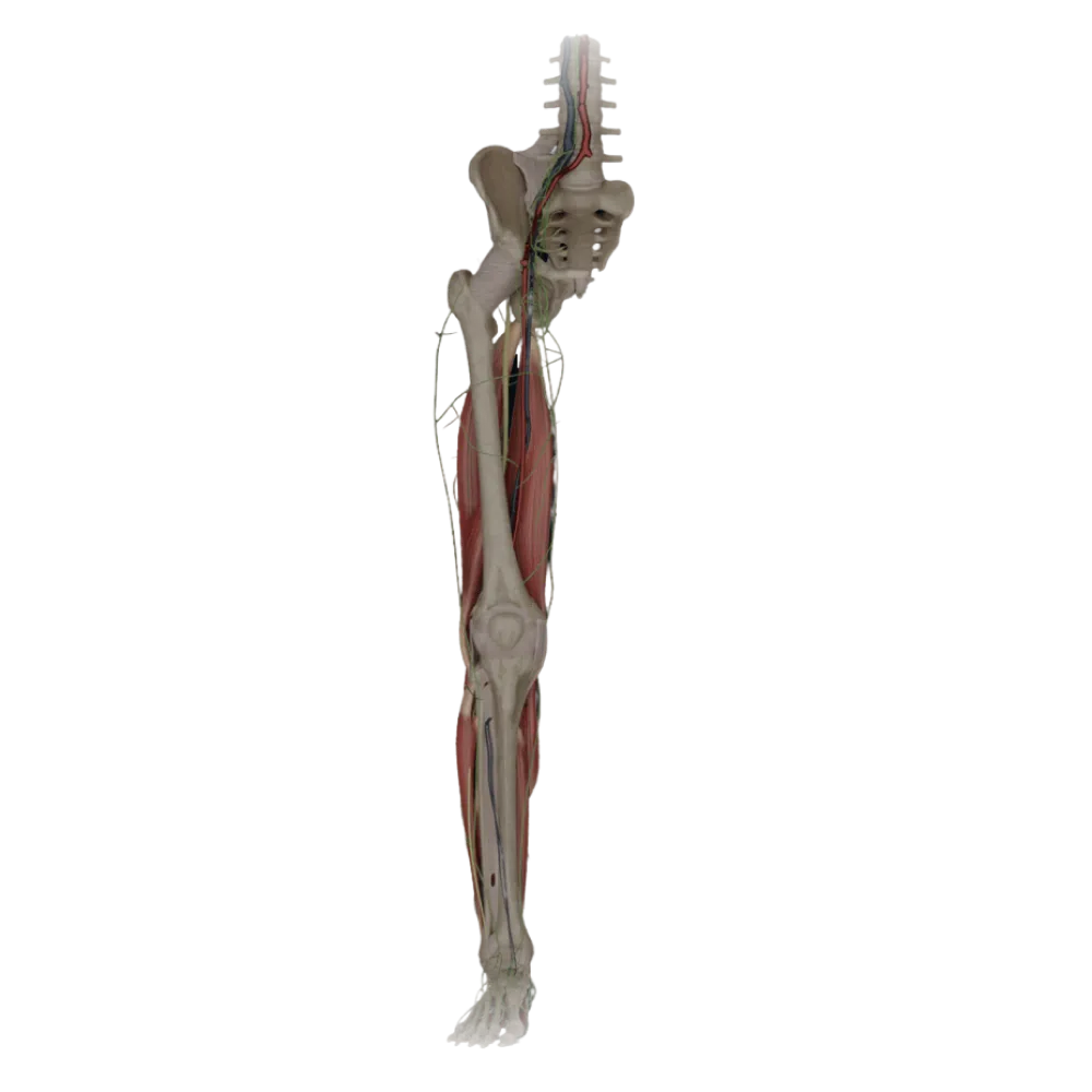
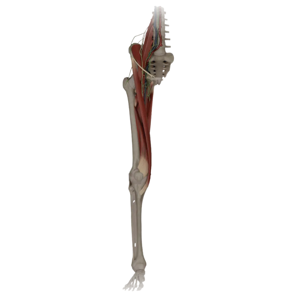
Thigh muscles anatomy test
Assess your knowledge of thigh muscle anatomy. The test examines their topography, blood supply sources, innervation zones, and crucial anastomoses.
1/20
bold
text
1. Which nerve innervates the quadriceps femoris muscle?
-
Obturator nerve.
The quadriceps femoris muscle belongs to the anterior group of thigh muscles and is innervated by motor branches of the femoral nerve (L2-L4).
-
Inferior gluteal nerve.
The quadriceps femoris muscle belongs to the anterior group of thigh muscles and is innervated by motor branches of the femoral nerve (L2-L4).
-
Sciatic nerve
The quadriceps femoris muscle belongs to the anterior group of thigh muscles and is innervated by motor branches of the femoral nerve (L2-L4).
-
Femoral nerve.
The quadriceps femoris muscle belongs to the anterior group of thigh muscles and is innervated by motor branches of the femoral nerve (L2-L4).
-
I find it difficult to answer
The quadriceps femoris muscle belongs to the anterior group of thigh muscles and is innervated by motor branches of the femoral nerve (L2-L4).
2. From what source are the muscles of the posterior thigh group primarily vascularized?
-
Obturator artery.
The primary source of blood supply for the posterior thigh muscle group (flexors) are the perforating arteries (aa. perforantes), branching from the deep femoral artery.
-
Descending genicular artery
The primary source of blood supply for the posterior thigh muscle group (flexors) are the perforating arteries (aa. perforantes), branching from the deep femoral artery.
-
Perforating arteries
The primary source of blood supply for the posterior thigh muscle group (flexors) are the perforating arteries (aa. perforantes), branching from the deep femoral artery.
-
External pudendal arteries
The primary source of blood supply for the posterior thigh muscle group (flexors) are the perforating arteries (aa. perforantes), branching from the deep femoral artery.
-
I find it difficult to answer
The primary source of blood supply for the posterior thigh muscle group (flexors) are the perforating arteries (aa. perforantes), branching from the deep femoral artery.
3. Which muscle of the medial thigh group has dual innervation (from the obturator and sciatic nerves)?
-
Adductor magnus muscle.
The adductor magnus muscle is innervated by the obturator nerve (its adducting part) and the tibial portion of the sciatic nerve (its extending part).
-
Gracilis muscle.
The adductor magnus muscle is innervated by the obturator nerve (its adducting part) and the tibial portion of the sciatic nerve (its extending part).
-
Pectineus muscle.
The adductor magnus muscle is innervated by the obturator nerve (its adducting part) and the tibial portion of the sciatic nerve (its extending part).
-
Adductor longus
The adductor magnus muscle is innervated by the obturator nerve (its adducting part) and the tibial portion of the sciatic nerve (its extending part).
-
I find it difficult to answer
The adductor magnus muscle is innervated by the obturator nerve (its adducting part) and the tibial portion of the sciatic nerve (its extending part).
4. Which artery branch supplies the vastus lateralis muscle?
-
Medial circumflex femoral artery
The vastus lateralis muscle is primarily supplied by the descending branch of the lateral circumflex femoral artery (ramus descendens a. circumflexae femoris lateralis).
-
Descending branch of the lateral circumflex femoral artery
The vastus lateralis muscle is primarily supplied by the descending branch of the lateral circumflex femoral artery (ramus descendens a. circumflexae femoris lateralis).
-
Obturator artery.
The vastus lateralis muscle is primarily supplied by the descending branch of the lateral circumflex femoral artery (ramus descendens a. circumflexae femoris lateralis).
-
Superficial circumflex iliac artery
The vastus lateralis muscle is primarily supplied by the descending branch of the lateral circumflex femoral artery (ramus descendens a. circumflexae femoris lateralis).
-
I find it difficult to answer
The vastus lateralis muscle is primarily supplied by the descending branch of the lateral circumflex femoral artery (ramus descendens a. circumflexae femoris lateralis).
5. Which part of the biceps femoris muscle is innervated by the common fibular nerve?
-
Long head
The short head of the biceps femoris is innervated by the common fibular portion of the sciatic nerve, unlike the long head, which is innervated by the tibial portion.
-
Both heads
The short head of the biceps femoris is innervated by the common fibular portion of the sciatic nerve, unlike the long head, which is innervated by the tibial portion.
-
Neither head
The short head of the biceps femoris is innervated by the common fibular portion of the sciatic nerve, unlike the long head, which is innervated by the tibial portion.
-
Short head
The short head of the biceps femoris is innervated by the common fibular portion of the sciatic nerve, unlike the long head, which is innervated by the tibial portion.
-
I find it difficult to answer
The short head of the biceps femoris is innervated by the common fibular portion of the sciatic nerve, unlike the long head, which is innervated by the tibial portion.
6. Which thigh muscle is primarily innervated by the femoral nerve but can receive additional branches from the obturator nerve?
-
Sartorius muscle.
Pectineus muscle is topographically and functionally transitional; its main innervation comes from the femoral nerve, with accessory innervation from the obturator.
-
Pectineus muscle.
Pectineus muscle is topographically and functionally transitional; its main innervation comes from the femoral nerve, with accessory innervation from the obturator.
-
Gracilis muscle.
Pectineus muscle is topographically and functionally transitional; its main innervation comes from the femoral nerve, with accessory innervation from the obturator.
-
Semitendinosus muscle.
Pectineus muscle is topographically and functionally transitional; its main innervation comes from the femoral nerve, with accessory innervation from the obturator.
-
I find it difficult to answer
Pectineus muscle is topographically and functionally transitional; its main innervation comes from the femoral nerve, with accessory innervation from the obturator.
7. Which artery passes through the adductor canal (canalis adductorius) together with the saphenous nerve?
-
Femoral artery.
The femoral artery passes through the adductor canal (Hunter's canal) in the thigh, accompanying the saphenous nerve, before it emerges in the popliteal fossa.
-
Deep femoral artery.
The femoral artery passes through the adductor canal (Hunter's canal) in the thigh, accompanying the saphenous nerve, before it emerges in the popliteal fossa.
-
Obturator artery.
The femoral artery passes through the adductor canal (Hunter's canal) in the thigh, accompanying the saphenous nerve, before it emerges in the popliteal fossa.
-
Popliteal artery.
The femoral artery passes through the adductor canal (Hunter's canal) in the thigh, accompanying the saphenous nerve, before it emerges in the popliteal fossa.
-
I find it difficult to answer
The femoral artery passes through the adductor canal (Hunter's canal) in the thigh, accompanying the saphenous nerve, before it emerges in the popliteal fossa.
8. Which of the listed muscles is NOT innervated by the obturator nerve?
-
Adductor brevis
The semimembranosus muscle belongs to the posterior group of thigh muscles and is innervated by the tibial portion of the sciatic nerve, not the obturator nerve.
-
Adductor longus
The semimembranosus muscle belongs to the posterior group of thigh muscles and is innervated by the tibial portion of the sciatic nerve, not the obturator nerve.
-
Semimembranosus muscle.
The semimembranosus muscle belongs to the posterior group of thigh muscles and is innervated by the tibial portion of the sciatic nerve, not the obturator nerve.
-
Gracilis muscle.
The semimembranosus muscle belongs to the posterior group of thigh muscles and is innervated by the tibial portion of the sciatic nerve, not the obturator nerve.
-
I find it difficult to answer
The semimembranosus muscle belongs to the posterior group of thigh muscles and is innervated by the tibial portion of the sciatic nerve, not the obturator nerve.
9. Which thigh muscle is pierced by the branches of the obturator nerve (dividing it into anterior and posterior branches)?
-
Pectineus muscle.
The obturator nerve divides into anterior and posterior branches, anatomically surrounding the short adductor muscle anteriorly and posteriorly, respectively.
-
Adductor magnus muscle.
The obturator nerve divides into anterior and posterior branches, anatomically surrounding the short adductor muscle anteriorly and posteriorly, respectively.
-
Gracilis muscle.
The obturator nerve divides into anterior and posterior branches, anatomically surrounding the short adductor muscle anteriorly and posteriorly, respectively.
-
Adductor brevis
The obturator nerve divides into anterior and posterior branches, anatomically surrounding the short adductor muscle anteriorly and posteriorly, respectively.
-
I find it difficult to answer
The obturator nerve divides into anterior and posterior branches, anatomically surrounding the short adductor muscle anteriorly and posteriorly, respectively.
10. The blood supply to the sartorius muscle (m. sartorius) is mainly provided by:
-
Perforating arteries
The sartorius muscle, located on the anterior surface of the thigh, receives numerous muscular branches (rr. musculares) directly from the trunk of the femoral artery.
-
Branches of the femoral artery
The sartorius muscle, located on the anterior surface of the thigh, receives numerous muscular branches (rr. musculares) directly from the trunk of the femoral artery.
-
The obturator artery
The sartorius muscle, located on the anterior surface of the thigh, receives numerous muscular branches (rr. musculares) directly from the trunk of the femoral artery.
-
The inferior gluteal artery
The sartorius muscle, located on the anterior surface of the thigh, receives numerous muscular branches (rr. musculares) directly from the trunk of the femoral artery.
-
I find it difficult to answer
The sartorius muscle, located on the anterior surface of the thigh, receives numerous muscular branches (rr. musculares) directly from the trunk of the femoral artery.
11. Which branch of the deep femoral artery participates in supplying blood to the muscles of the medial group and anastomoses with the obturator artery?
-
Medial circumflex femoral artery
The medial artery, circling the femur, actively supplies blood to the adductor muscles and forms essential anastomoses with the branches of the obturator artery.
-
Lateral circumflex femoral artery
The medial artery, circling the femur, actively supplies blood to the adductor muscles and forms essential anastomoses with the branches of the obturator artery.
-
First perforating artery
The medial artery, circling the femur, actively supplies blood to the adductor muscles and forms essential anastomoses with the branches of the obturator artery.
-
Descending genicular artery
The medial artery, circling the femur, actively supplies blood to the adductor muscles and forms essential anastomoses with the branches of the obturator artery.
-
I find it difficult to answer
The medial artery, circling the femur, actively supplies blood to the adductor muscles and forms essential anastomoses with the branches of the obturator artery.
12. Which nerve innervates the long head of the biceps femoris muscle?
-
Femoral nerve
The long head of the biceps femoris muscle is innervated by the tibial part of the sciatic nerve, like other typical flexors of the posterior thigh group.
-
Obturator nerve
The long head of the biceps femoris muscle is innervated by the tibial part of the sciatic nerve, like other typical flexors of the posterior thigh group.
-
Tibial nerve (portion of the sciatic)
The long head of the biceps femoris muscle is innervated by the tibial part of the sciatic nerve, like other typical flexors of the posterior thigh group.
-
Common fibular nerve
The long head of the biceps femoris muscle is innervated by the tibial part of the sciatic nerve, like other typical flexors of the posterior thigh group.
-
I find it difficult to answer
The long head of the biceps femoris muscle is innervated by the tibial part of the sciatic nerve, like other typical flexors of the posterior thigh group.
13. Which of the listed muscles receives arterial blood from a. genus descendens (descending genicular artery)?
-
Biceps femoris muscle.
The descending genicular artery branches from the femoral artery in the adductor canal, perforates its medial wall, and supplies the vastus medialis muscle.
-
Adductor brevis
The descending genicular artery branches from the femoral artery in the adductor canal, perforates its medial wall, and supplies the vastus medialis muscle.
-
Quadratus femoris muscle.
The descending genicular artery branches from the femoral artery in the adductor canal, perforates its medial wall, and supplies the vastus medialis muscle.
-
Vastus medialis
The descending genicular artery branches from the femoral artery in the adductor canal, perforates its medial wall, and supplies the vastus medialis muscle.
-
I find it difficult to answer
The descending genicular artery branches from the femoral artery in the adductor canal, perforates its medial wall, and supplies the vastus medialis muscle.
14. What structure innervates the muscle that tenses the fascia lata (m. tensor fasciae latae)?
-
Superior gluteal nerve
Despite its location on the anterolateral surface of the thigh, the muscle tensing the fascia lata develops from the gluteal plat and is innervated by the superior gluteal nerve.
-
Femoral nerve.
Despite its location on the anterolateral surface of the thigh, the muscle tensing the fascia lata develops from the gluteal plat and is innervated by the superior gluteal nerve.
-
Inferior gluteal nerve.
Despite its location on the anterolateral surface of the thigh, the muscle tensing the fascia lata develops from the gluteal plat and is innervated by the superior gluteal nerve.
-
Lateral femoral cutaneous nerve.
Despite its location on the anterolateral surface of the thigh, the muscle tensing the fascia lata develops from the gluteal plat and is innervated by the superior gluteal nerve.
-
I find it difficult to answer
Despite its location on the anterolateral surface of the thigh, the muscle tensing the fascia lata develops from the gluteal plat and is innervated by the superior gluteal nerve.
15. Through which opening do the obturator artery and nerve exit to the medial thigh for supplying blood and innervation to the muscles?
-
Suprapiriform foramen
The obturator neurovascular bundle exits the pelvic cavity and enters the medial (adductor) compartment of the thigh through the name-sake obturator canal.
-
Infrapiriform foramen
The obturator neurovascular bundle exits the pelvic cavity and enters the medial (adductor) compartment of the thigh through the name-sake obturator canal.
-
Obturator canal.
The obturator neurovascular bundle exits the pelvic cavity and enters the medial (adductor) compartment of the thigh through the name-sake obturator canal.
-
Femoral canal
The obturator neurovascular bundle exits the pelvic cavity and enters the medial (adductor) compartment of the thigh through the name-sake obturator canal.
-
I find it difficult to answer
The obturator neurovascular bundle exits the pelvic cavity and enters the medial (adductor) compartment of the thigh through the name-sake obturator canal.
16. Which arteries form the so-called cruciate network (anastomosis) of the thigh, providing collateral circulation?
-
Femoral, popliteal, and obturator arteries
The cruciate anastomosis on the posterior surface of the thigh is formed by branches of the a. Circumflexa femoris medialis et lateralis, a. glutea inferior and a. Perforans prima.
-
Medial and lateral circumflex femoral arteries, the inferior gluteal artery, and the 1st perforating artery.
The cruciate anastomosis on the posterior surface of the thigh is formed by branches of the a. Circumflexa femoris medialis et lateralis, a. glutea inferior and a. Perforans prima.
-
External and internal pudendal arteries
The cruciate anastomosis on the posterior surface of the thigh is formed by branches of the a. Circumflexa femoris medialis et lateralis, a. glutea inferior and a. Perforans prima.
-
Deep femoral artery and descending genicular artery
The cruciate anastomosis on the posterior surface of the thigh is formed by branches of the a. Circumflexa femoris medialis et lateralis, a. glutea inferior and a. Perforans prima.
-
I find it difficult to answer
The cruciate anastomosis on the posterior surface of the thigh is formed by branches of the a. Circumflexa femoris medialis et lateralis, a. glutea inferior and a. Perforans prima.
17. Which thigh muscle forms the lateral wall of the femoral triangle and is innervated by the femoral nerve?
-
Gracilis muscle.
The sartorius muscle (m. sartorius) constitutes the lateral boundary of the femoral triangle (Scarpa's) and consistently receives innervation from the n. Femoralis.
-
Adductor longus
The sartorius muscle (m. sartorius) constitutes the lateral boundary of the femoral triangle (Scarpa's) and consistently receives innervation from the n. Femoralis.
-
Pectineus muscle.
The sartorius muscle (m. sartorius) constitutes the lateral boundary of the femoral triangle (Scarpa's) and consistently receives innervation from the n. Femoralis.
-
Sartorius muscle.
The sartorius muscle (m. sartorius) constitutes the lateral boundary of the femoral triangle (Scarpa's) and consistently receives innervation from the n. Femoralis.
-
I find it difficult to answer
The sartorius muscle (m. sartorius) constitutes the lateral boundary of the femoral triangle (Scarpa's) and consistently receives innervation from the n. Femoralis.
18. Which artery is the primary source of blood supply for the deep structures of the thigh and branches from the femoral artery in the femoral triangle?
-
Deep femoral artery.
Deep femoral artery (a. profunda femoris) is the largest and functionally significant branch of the femoral artery, supplying the majority of the thigh muscles with blood.
-
Obturator artery.
Deep femoral artery (a. profunda femoris) is the largest and functionally significant branch of the femoral artery, supplying the majority of the thigh muscles with blood.
-
External iliac artery
Deep femoral artery (a. profunda femoris) is the largest and functionally significant branch of the femoral artery, supplying the majority of the thigh muscles with blood.
-
Superficial epigastric artery
Deep femoral artery (a. profunda femoris) is the largest and functionally significant branch of the femoral artery, supplying the majority of the thigh muscles with blood.
-
I find it difficult to answer
Deep femoral artery (a. profunda femoris) is the largest and functionally significant branch of the femoral artery, supplying the majority of the thigh muscles with blood.
19. Which muscle of the posterior thigh group attaches to the head of the fibula and receives dual innervation from different parts of the sciatic nerve?
-
Semimembranosus muscle.
The biceps femoris muscle has a long head (tibial portion) and a short head (common fibular portion), and its tendon attaches to the fibula.
-
Semitendinosus muscle.
The biceps femoris muscle has a long head (tibial portion) and a short head (common fibular portion), and its tendon attaches to the fibula.
-
Biceps femoris muscle.
The biceps femoris muscle has a long head (tibial portion) and a short head (common fibular portion), and its tendon attaches to the fibula.
-
Adductor magnus muscle.
The biceps femoris muscle has a long head (tibial portion) and a short head (common fibular portion), and its tendon attaches to the fibula.
-
I find it difficult to answer
The biceps femoris muscle has a long head (tibial portion) and a short head (common fibular portion), and its tendon attaches to the fibula.
20. Which branch of the femoral nerve is the longest, purely sensory in nature, and accompanies the femoral artery in the adductor canal?
-
Lateral femoral cutaneous nerve.
The saphenous nerve (n. saphenus) is the longest sensory branch of the femoral nerve; it does not innervate muscles but is topographically closely associated with femoral vessels.
-
Saphenous nerve (n. saphenus)
The saphenous nerve (n. saphenus) is the longest sensory branch of the femoral nerve; it does not innervate muscles but is topographically closely associated with femoral vessels.
-
Posterior femoral cutaneous nerve.
The saphenous nerve (n. saphenus) is the longest sensory branch of the femoral nerve; it does not innervate muscles but is topographically closely associated with femoral vessels.
-
Muscular branch to the sartorius muscle
The saphenous nerve (n. saphenus) is the longest sensory branch of the femoral nerve; it does not innervate muscles but is topographically closely associated with femoral vessels.
-
I find it difficult to answer
The saphenous nerve (n. saphenus) is the longest sensory branch of the femoral nerve; it does not innervate muscles but is topographically closely associated with femoral vessels.
Retake this quiz?
Your current progress will be reset.