1/20
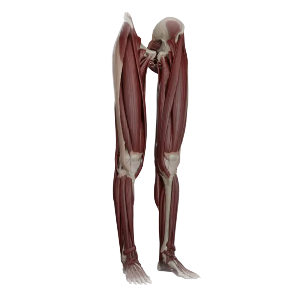

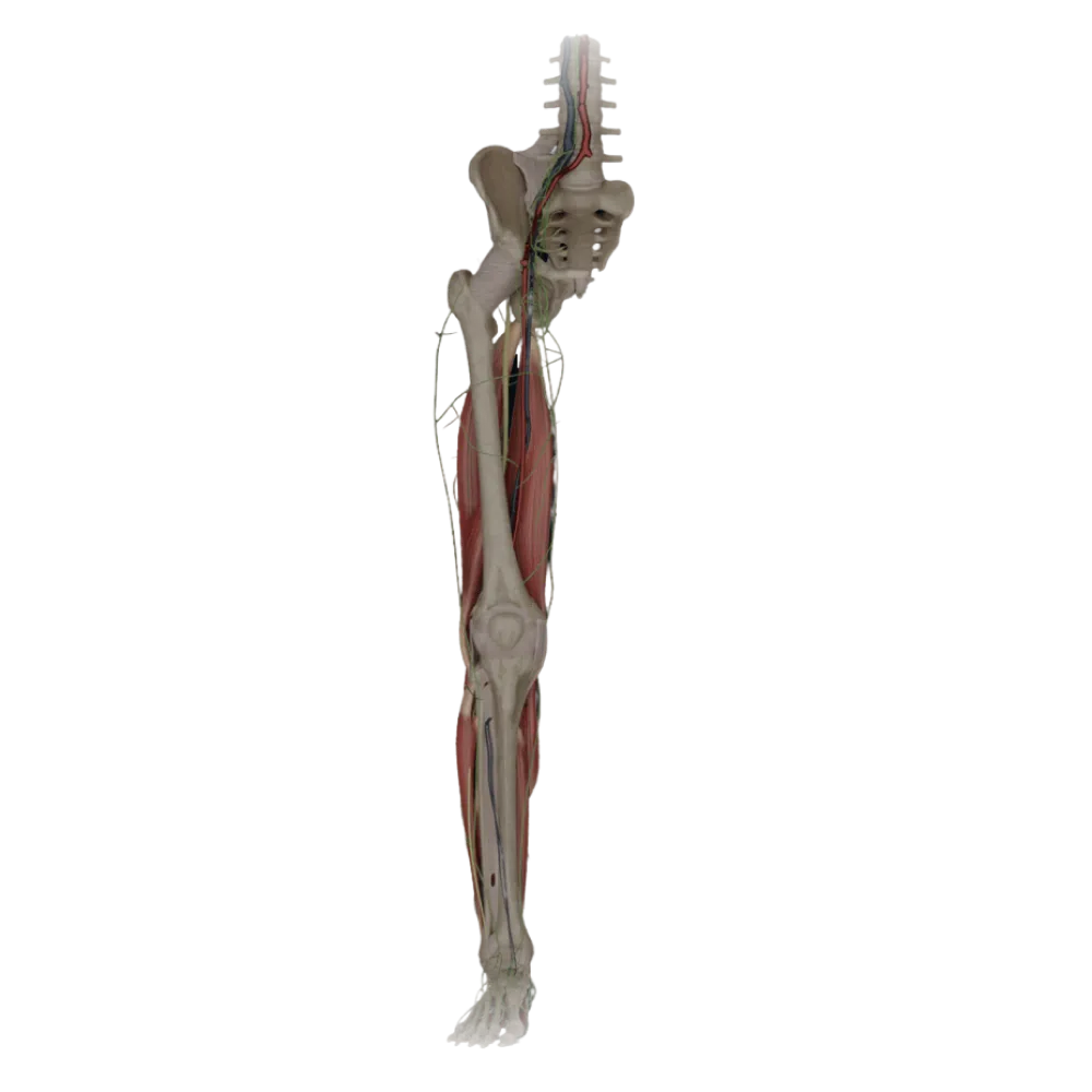
Anatomy test of lower limb muscles.
Test your knowledge of myology of the lower limb. The test covers topography, functions, attachment points, and fascial channels of leg muscles.
1. Which muscle exits the pelvic cavity through the greater sciatic foramen?
-
Piriformis muscle (M. piriformis).
The piriformis muscle passes through the greater sciatic foramen, dividing it into the suprapiriform and infrapiriform foramina.
-
Obturator internus muscle (M. obturatorius internus).
The piriformis muscle passes through the greater sciatic foramen, dividing it into the suprapiriform and infrapiriform foramina.
-
Obturator externus muscle (M. obturatorius externus).
The piriformis muscle passes through the greater sciatic foramen, dividing it into the suprapiriform and infrapiriform foramina.
-
Quadratus femoris muscle (M. quadratus femoris).
The piriformis muscle passes through the greater sciatic foramen, dividing it into the suprapiriform and infrapiriform foramina.
-
I find it difficult to answer
The piriformis muscle passes through the greater sciatic foramen, dividing it into the suprapiriform and infrapiriform foramina.
2. To which muscle group does the pectineus muscle (m. pectineus) belong?
-
Anterior group of thigh muscles.
The pectineus muscle belongs to the medial group of thigh muscles, primarily performing the function of adduction.
-
Medial group of thigh muscles.
The pectineus muscle belongs to the medial group of thigh muscles, primarily performing the function of adduction.
-
Posterior group of thigh muscles.
The pectineus muscle belongs to the medial group of thigh muscles, primarily performing the function of adduction.
-
Internal group of pelvic muscles.
The pectineus muscle belongs to the medial group of thigh muscles, primarily performing the function of adduction.
-
I find it difficult to answer
The pectineus muscle belongs to the medial group of thigh muscles, primarily performing the function of adduction.
3. Which muscle forms the floor of the popliteal fossa?
-
Biceps femoris muscle.
The floor of the popliteal fossa is formed by the popliteal surface of the femur, the joint capsule of the knee, and the popliteus muscle.
-
Gastrocnemius muscle.
The floor of the popliteal fossa is formed by the popliteal surface of the femur, the joint capsule of the knee, and the popliteus muscle.
-
Popliteus muscle (M. popliteus).
The floor of the popliteal fossa is formed by the popliteal surface of the femur, the joint capsule of the knee, and the popliteus muscle.
-
Semimembranosus muscle.
The floor of the popliteal fossa is formed by the popliteal surface of the femur, the joint capsule of the knee, and the popliteus muscle.
-
I find it difficult to answer
The floor of the popliteal fossa is formed by the popliteal surface of the femur, the joint capsule of the knee, and the popliteus muscle.
4. Which muscle does NOT attach to the pes anserinus (goose's foot)?
-
Sartorius muscle (M. sartorius).
The tendons of the sartorius, gracilis, and semitendinosus muscles attach to the superficial pes anserinus. The semimembranosus muscle forms the deep pes anserinus.
-
Gracilis muscle (M. gracilis).
The tendons of the sartorius, gracilis, and semitendinosus muscles attach to the superficial pes anserinus. The semimembranosus muscle forms the deep pes anserinus.
-
Semitendinosus muscle (M. semitendinosus).
The tendons of the sartorius, gracilis, and semitendinosus muscles attach to the superficial pes anserinus. The semimembranosus muscle forms the deep pes anserinus.
-
Semimembranosus muscle (M. semimembranosus).
The tendons of the sartorius, gracilis, and semitendinosus muscles attach to the superficial pes anserinus. The semimembranosus muscle forms the deep pes anserinus.
-
I find it difficult to answer
The tendons of the sartorius, gracilis, and semitendinosus muscles attach to the superficial pes anserinus. The semimembranosus muscle forms the deep pes anserinus.
5. Which structure limits the femoral canal laterally?
-
Femoral vein
The femoral canal is limited laterally by the wall of the femoral vein.
-
Lacunar ligament.
The femoral canal is limited laterally by the wall of the femoral vein.
-
Inguinal ligament
The femoral canal is limited laterally by the wall of the femoral vein.
-
Pectineal ligament.
The femoral canal is limited laterally by the wall of the femoral vein.
-
I find it difficult to answer
The femoral canal is limited laterally by the wall of the femoral vein.
6. Identify the muscle whose tendon passes in a separate synovial sheath beneath the flexor retinaculum (retinaculum musculorum flexorum) behind the medial malleolus, nearest to it:
-
Flexor digitorum longus
Behind the medial malleolus, the tendon of the posterior tibial muscle is the first to pass (most medially and anteriorly).
-
Tibialis posterior muscle.
Behind the medial malleolus, the tendon of the posterior tibial muscle is the first to pass (most medially and anteriorly).
-
Flexor hallucis longus.
Behind the medial malleolus, the tendon of the posterior tibial muscle is the first to pass (most medially and anteriorly).
-
Tibialis anterior muscle.
Behind the medial malleolus, the tendon of the posterior tibial muscle is the first to pass (most medially and anteriorly).
-
I find it difficult to answer
Behind the medial malleolus, the tendon of the posterior tibial muscle is the first to pass (most medially and anteriorly).
7. Which of the listed muscles flexes the thigh and extends the leg?
-
Semitendinosus muscle (M. semitendinosus).
The rectus femoris muscle is biarticular: it crosses both the hip (flexes) and knee (extends) joints.
-
Biceps femoris muscle (M. biceps femoris).
The rectus femoris muscle is biarticular: it crosses both the hip (flexes) and knee (extends) joints.
-
Rectus femoris muscle (M. rectus femoris).
The rectus femoris muscle is biarticular: it crosses both the hip (flexes) and knee (extends) joints.
-
Sartorius muscle (M. sartorius).
The rectus femoris muscle is biarticular: it crosses both the hip (flexes) and knee (extends) joints.
-
I find it difficult to answer
The rectus femoris muscle is biarticular: it crosses both the hip (flexes) and knee (extends) joints.
8. Which muscle of the leg performs plantar flexion, supination, and adduction of the foot?
-
Fibularis longus muscle.
The posterior tibial muscle is the main supinator and adducting muscle of the foot and also participates in plantar flexion.
-
Tibialis anterior muscle.
The posterior tibial muscle is the main supinator and adducting muscle of the foot and also participates in plantar flexion.
-
Fibularis brevis muscle.
The posterior tibial muscle is the main supinator and adducting muscle of the foot and also participates in plantar flexion.
-
Tibialis posterior muscle.
The posterior tibial muscle is the main supinator and adducting muscle of the foot and also participates in plantar flexion.
-
I find it difficult to answer
The posterior tibial muscle is the main supinator and adducting muscle of the foot and also participates in plantar flexion.
9. Through which opening does the obturator nerve exit to the thigh?
-
Obturator canal.
The obturator nerve, along with the homonymous vessels, exits to the thigh through the obturator canal (canalis obturatorius).
-
Muscular lacuna.
The obturator nerve, along with the homonymous vessels, exits to the thigh through the obturator canal (canalis obturatorius).
-
Vascular lacuna.
The obturator nerve, along with the homonymous vessels, exits to the thigh through the obturator canal (canalis obturatorius).
-
Suprapiriform foramen
The obturator nerve, along with the homonymous vessels, exits to the thigh through the obturator canal (canalis obturatorius).
-
I find it difficult to answer
The obturator nerve, along with the homonymous vessels, exits to the thigh through the obturator canal (canalis obturatorius).
10. What forms the anterior wall of the adductor (Hunter's) canal?
-
Adductor magnus muscle.
The anterior wall of the adductor canal is a fibrous plate stretched between the vastus medialis and the adductor magnus muscles.
-
Adductor longus muscle.
The anterior wall of the adductor canal is a fibrous plate stretched between the vastus medialis and the adductor magnus muscles.
-
Vastus medialis muscle of the thigh.
The anterior wall of the adductor canal is a fibrous plate stretched between the vastus medialis and the adductor magnus muscles.
-
Fibrous plate (lamina vastoadductoria).
The anterior wall of the adductor canal is a fibrous plate stretched between the vastus medialis and the adductor magnus muscles.
-
I find it difficult to answer
The anterior wall of the adductor canal is a fibrous plate stretched between the vastus medialis and the adductor magnus muscles.
11. Identify the muscle that does NOT belong to the deep layer of the posterior group of calf muscles:
-
Tibialis posterior muscle.
The soleus muscle, along with the gastrocnemius, forms the triceps surae, which belongs to the superficial layer.
-
Flexor digitorum longus
The soleus muscle, along with the gastrocnemius, forms the triceps surae, which belongs to the superficial layer.
-
Soleus muscle.
The soleus muscle, along with the gastrocnemius, forms the triceps surae, which belongs to the superficial layer.
-
Flexor hallucis longus.
The soleus muscle, along with the gastrocnemius, forms the triceps surae, which belongs to the superficial layer.
-
I find it difficult to answer
The soleus muscle, along with the gastrocnemius, forms the triceps surae, which belongs to the superficial layer.
12. Which muscle attaches to the tuberosity of the fifth metatarsal bone?
-
Fibularis longus muscle.
The tendon of the fibularis brevis muscle attaches to the tuberosity of the fifth metatarsal bone.
-
Fibularis brevis muscle.
The tendon of the fibularis brevis muscle attaches to the tuberosity of the fifth metatarsal bone.
-
Fibularis tertius muscle.
The tendon of the fibularis brevis muscle attaches to the tuberosity of the fifth metatarsal bone.
-
Tibialis posterior muscle.
The tendon of the fibularis brevis muscle attaches to the tuberosity of the fifth metatarsal bone.
-
I find it difficult to answer
The tendon of the fibularis brevis muscle attaches to the tuberosity of the fifth metatarsal bone.
13. Which of the pelvic muscles originates on the pelvic surface of the sacrum?
-
Piriformis muscle.
The piriformis muscle originates on the pelvic surface of the sacrum lateral to the pelvic sacral foramina.
-
Obturator internus muscle.
The piriformis muscle originates on the pelvic surface of the sacrum lateral to the pelvic sacral foramina.
-
Obturator externus muscle.
The piriformis muscle originates on the pelvic surface of the sacrum lateral to the pelvic sacral foramina.
-
Quadratus femoris muscle.
The piriformis muscle originates on the pelvic surface of the sacrum lateral to the pelvic sacral foramina.
-
I find it difficult to answer
The piriformis muscle originates on the pelvic surface of the sacrum lateral to the pelvic sacral foramina.
14. What forms the superior border of the femoral triangle (of Scarpa)?
-
Sartorius muscle.
The femoral triangle is bounded superiorly by the inguinal ligament, laterally by the sartorius muscle, and medially by the adductor longus muscle.
-
Adductor longus
The femoral triangle is bounded superiorly by the inguinal ligament, laterally by the sartorius muscle, and medially by the adductor longus muscle.
-
Inguinal ligament
The femoral triangle is bounded superiorly by the inguinal ligament, laterally by the sartorius muscle, and medially by the adductor longus muscle.
-
Pectineus muscle.
The femoral triangle is bounded superiorly by the inguinal ligament, laterally by the sartorius muscle, and medially by the adductor longus muscle.
-
I find it difficult to answer
The femoral triangle is bounded superiorly by the inguinal ligament, laterally by the sartorius muscle, and medially by the adductor longus muscle.
15. Which muscle is located within the vascular lacuna (lacuna vasorum)?
-
Iliopsoas muscle
Within the vascular lacuna pass the femoral artery and vein as well as the Rosenmüller-Pirogov lymph node. The iliopsoas muscle passes through the muscular lacuna.
-
Pectineus muscle.
Within the vascular lacuna pass the femoral artery and vein as well as the Rosenmüller-Pirogov lymph node. The iliopsoas muscle passes through the muscular lacuna.
-
Tensor fasciae latae
Within the vascular lacuna pass the femoral artery and vein as well as the Rosenmüller-Pirogov lymph node. The iliopsoas muscle passes through the muscular lacuna.
-
No muscle is located there.
Within the vascular lacuna pass the femoral artery and vein as well as the Rosenmüller-Pirogov lymph node. The iliopsoas muscle passes through the muscular lacuna.
-
I find it difficult to answer
Within the vascular lacuna pass the femoral artery and vein as well as the Rosenmüller-Pirogov lymph node. The iliopsoas muscle passes through the muscular lacuna.
16. Indicate the function of the tensor fasciae latae:
-
Tenses the fascia lata, flexes, abducts, and pronates the thigh.
The tensor fasciae latae assists in flexion, abduction, and internal rotation (pronation) of the thigh, while tensing the iliotibial tract.
-
Extends and supinates the thigh.
The tensor fasciae latae assists in flexion, abduction, and internal rotation (pronation) of the thigh, while tensing the iliotibial tract.
-
Adducts the thigh and flexes the leg.
The tensor fasciae latae assists in flexion, abduction, and internal rotation (pronation) of the thigh, while tensing the iliotibial tract.
-
Tenses the fascia lata and extends the knee joint.
The tensor fasciae latae assists in flexion, abduction, and internal rotation (pronation) of the thigh, while tensing the iliotibial tract.
-
I find it difficult to answer
The tensor fasciae latae assists in flexion, abduction, and internal rotation (pronation) of the thigh, while tensing the iliotibial tract.
17. Which foot muscle is located in the medial group of plantar muscles?
-
Abductor digiti minimi muscle.
The abductor hallucis muscle belongs to the medial group (muscles of the great toe eminence).
-
Flexor digitorum brevis muscle.
The abductor hallucis muscle belongs to the medial group (muscles of the great toe eminence).
-
Abductor hallucis muscle.
The abductor hallucis muscle belongs to the medial group (muscles of the great toe eminence).
-
Quadratus plantae muscle.
The abductor hallucis muscle belongs to the medial group (muscles of the great toe eminence).
-
I find it difficult to answer
The abductor hallucis muscle belongs to the medial group (muscles of the great toe eminence).
18. What anatomical structure lies between the superficial and deep layers of the posterior group of calf muscles?
-
Superior muscular-peroneal canal.
Gruber's cruro-popliteal canal is located between the soleus muscle (superficial layer) and the deep muscles of the posterior group of the calf.
-
Cruripoplietal canal (canalis cruropopliteus).
Gruber's cruro-popliteal canal is located between the soleus muscle (superficial layer) and the deep muscles of the posterior group of the calf.
-
Inferior muscular-peroneal canal.
Gruber's cruro-popliteal canal is located between the soleus muscle (superficial layer) and the deep muscles of the posterior group of the calf.
-
Adductor canal.
Gruber's cruro-popliteal canal is located between the soleus muscle (superficial layer) and the deep muscles of the posterior group of the calf.
-
I find it difficult to answer
Gruber's cruro-popliteal canal is located between the soleus muscle (superficial layer) and the deep muscles of the posterior group of the calf.
19. Which muscle originates from the anterior superior iliac spine (spina iliaca anterior superior)?
-
Rectus femoris.
The sartorius muscle and tensor fasciae latae originate from the spina iliaca anterior superior. The rectus femoris muscle originates from the spina iliaca anterior inferior.
-
Gracilis muscle.
The sartorius muscle and tensor fasciae latae originate from the spina iliaca anterior superior. The rectus femoris muscle originates from the spina iliaca anterior inferior.
-
Iliacus muscle.
The sartorius muscle and tensor fasciae latae originate from the spina iliaca anterior superior. The rectus femoris muscle originates from the spina iliaca anterior inferior.
-
Sartorius muscle (M. sartorius).
The sartorius muscle and tensor fasciae latae originate from the spina iliaca anterior superior. The rectus femoris muscle originates from the spina iliaca anterior inferior.
-
I find it difficult to answer
The sartorius muscle and tensor fasciae latae originate from the spina iliaca anterior superior. The rectus femoris muscle originates from the spina iliaca anterior inferior.
20. Which muscle is the most powerful muscle extending the hip joint?
-
Gluteus medius muscle.
The gluteus maximus muscle is the main and most powerful extensor of the thigh in the hip joint.
-
Gluteus maximus muscle (M. gluteus maximus).
The gluteus maximus muscle is the main and most powerful extensor of the thigh in the hip joint.
-
Biceps femoris muscle.
The gluteus maximus muscle is the main and most powerful extensor of the thigh in the hip joint.
-
Adductor magnus muscle.
The gluteus maximus muscle is the main and most powerful extensor of the thigh in the hip joint.
-
I find it difficult to answer
The gluteus maximus muscle is the main and most powerful extensor of the thigh in the hip joint.
Retake this quiz?
Your current progress will be reset.