1/20
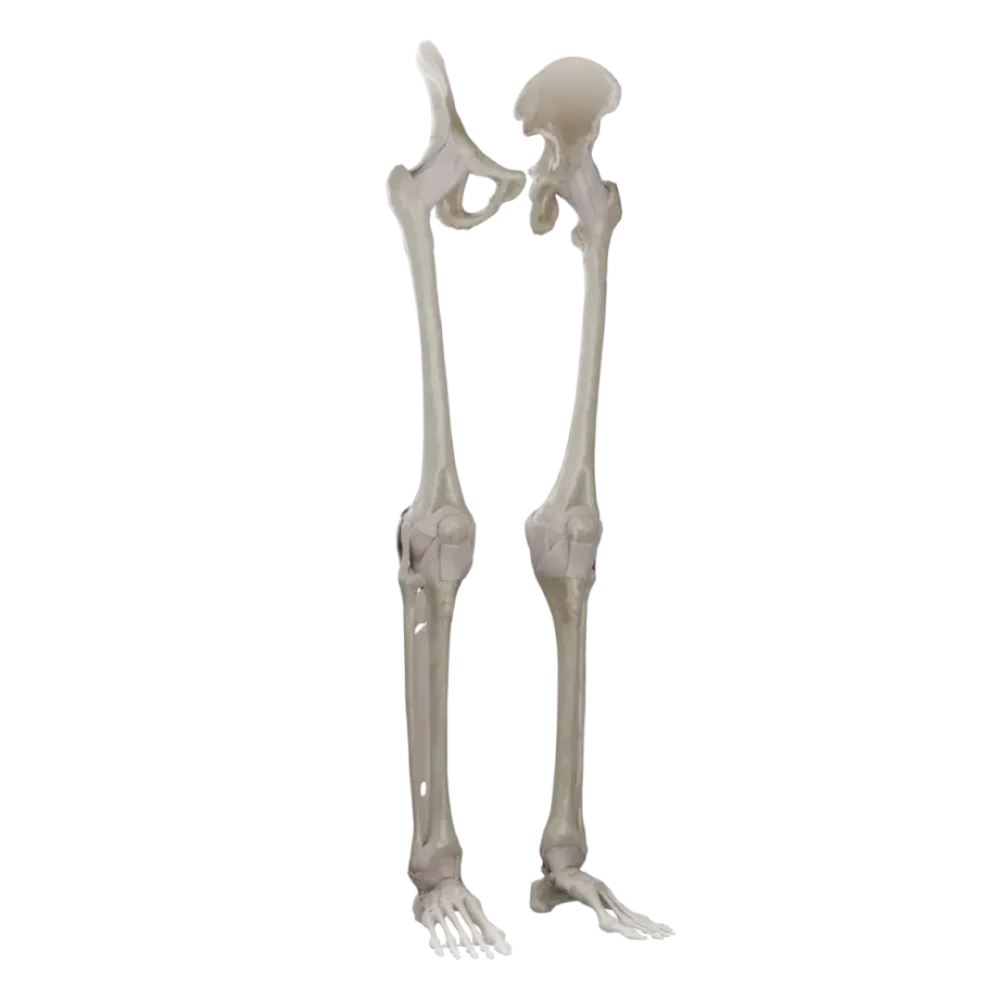

Anatomy test of joints of lower limb
Evaluate the knowledge of joints of lower limb anatomy. The test examines the topography and biomechanics of the joints, ligaments, and menisci of the pelvis and leg.
1. Which ligament of the hip joint is the strongest and inhibits extension?
-
Iliofemoral
The iliofemoral ligament (lig. iliofemorale) is the strongest in the body, preventing extension in the hip joint.
-
Circular zone
The iliofemoral ligament (lig. iliofemorale) is the strongest in the body, preventing extension in the hip joint.
-
Pubofemoral
The iliofemoral ligament (lig. iliofemorale) is the strongest in the body, preventing extension in the hip joint.
-
Ischiofemoral
The iliofemoral ligament (lig. iliofemorale) is the strongest in the body, preventing extension in the hip joint.
-
I find it difficult to answer
The iliofemoral ligament (lig. iliofemorale) is the strongest in the body, preventing extension in the hip joint.
2. Which structure inside the knee joint connects the anterior intercondylar area of the tibia with the lateral condyle of the femur?
-
Posterior cruciate ligament
The anterior cruciate ligament originates from the medial surface of the lateral femoral condyle and attaches to the area intercondylaris anterior.
-
Anterior cruciate ligament
The anterior cruciate ligament originates from the medial surface of the lateral femoral condyle and attaches to the area intercondylaris anterior.
-
Transverse ligament of knee
The anterior cruciate ligament originates from the medial surface of the lateral femoral condyle and attaches to the area intercondylaris anterior.
-
Medial meniscus
The anterior cruciate ligament originates from the medial surface of the lateral femoral condyle and attaches to the area intercondylaris anterior.
-
I find it difficult to answer
The anterior cruciate ligament originates from the medial surface of the lateral femoral condyle and attaches to the area intercondylaris anterior.
3. Which ligament is not part of the ankle (talocrural) joint ligaments?
-
Medial (deltoid) ligament
The bifurcate ligament (lig. bifurcatum) is the key to the transverse tarsal joint (Chopart's joint), not the ankle joint.
-
Anterior talofibular ligament.
The bifurcate ligament (lig. bifurcatum) is the key to the transverse tarsal joint (Chopart's joint), not the ankle joint.
-
Bifurcate ligament.
The bifurcate ligament (lig. bifurcatum) is the key to the transverse tarsal joint (Chopart's joint), not the ankle joint.
-
Calcaneofibular ligament.
The bifurcate ligament (lig. bifurcatum) is the key to the transverse tarsal joint (Chopart's joint), not the ankle joint.
-
I find it difficult to answer
The bifurcate ligament (lig. bifurcatum) is the key to the transverse tarsal joint (Chopart's joint), not the ankle joint.
4. Which bones form the transverse tarsal joint (Chopart's joint)?
-
Talus, calcaneus, navicular, and cuboid
The transverse tarsal joint is formed by two joints: the talonavicular and calcaneocuboid joints.
-
Talus, calcaneus, and cuneiforms
The transverse tarsal joint is formed by two joints: the talonavicular and calcaneocuboid joints.
-
Navicular, cuboid, and cuneiforms
The transverse tarsal joint is formed by two joints: the talonavicular and calcaneocuboid joints.
-
Calcaneus, cuboid, and metatarsals
The transverse tarsal joint is formed by two joints: the talonavicular and calcaneocuboid joints.
-
I find it difficult to answer
The transverse tarsal joint is formed by two joints: the talonavicular and calcaneocuboid joints.
5. Which ligament firmly connects the heads of the metatarsals, supporting the transverse arch of the foot?
-
Long plantar ligament.
The deep transverse metatarsal ligament connects the heads of the metatarsals, reinforcing the transverse arch of the foot.
-
Plantar aponeurosis.
The deep transverse metatarsal ligament connects the heads of the metatarsals, reinforcing the transverse arch of the foot.
-
Deep transverse metatarsal ligament
The deep transverse metatarsal ligament connects the heads of the metatarsals, reinforcing the transverse arch of the foot.
-
Short plantar ligament.
The deep transverse metatarsal ligament connects the heads of the metatarsals, reinforcing the transverse arch of the foot.
-
I find it difficult to answer
The deep transverse metatarsal ligament connects the heads of the metatarsals, reinforcing the transverse arch of the foot.
6. Which intraarticular structure is located in the cavity of the hip joint?
-
Transverse acetabular ligament
The ligament of the head of the femur (lig. capitis femoris) is located inside the joint and contains blood vessels to the head of the femur.
-
Ligament of head of femur.
The ligament of the head of the femur (lig. capitis femoris) is located inside the joint and contains blood vessels to the head of the femur.
-
Circular zone
The ligament of the head of the femur (lig. capitis femoris) is located inside the joint and contains blood vessels to the head of the femur.
-
Iliofemoral ligament.
The ligament of the head of the femur (lig. capitis femoris) is located inside the joint and contains blood vessels to the head of the femur.
-
I find it difficult to answer
The ligament of the head of the femur (lig. capitis femoris) is located inside the joint and contains blood vessels to the head of the femur.
7. Which structure is traditionally considered the key to the Lisfranc joint (tarsometatarsal joints)?
-
Bifurcate ligament.
The medial cuneometatarsal interosseous ligament (lig. cuneometatarsale interosseum mediale) firmly secures the Lisfranc joint.
-
Long plantar ligament.
The medial cuneometatarsal interosseous ligament (lig. cuneometatarsale interosseum mediale) firmly secures the Lisfranc joint.
-
Medial cuneometatarsal interosseous ligament
The medial cuneometatarsal interosseous ligament (lig. cuneometatarsale interosseum mediale) firmly secures the Lisfranc joint.
-
Plantar calcaneonavicular ligament.
The medial cuneometatarsal interosseous ligament (lig. cuneometatarsale interosseum mediale) firmly secures the Lisfranc joint.
-
I find it difficult to answer
The medial cuneometatarsal interosseous ligament (lig. cuneometatarsale interosseum mediale) firmly secures the Lisfranc joint.
8. With which structures does the knee joint capsule firmly adhere on its medial side?
-
With the fibular collateral ligament
The capsule of the knee joint tightly adheres to the medial collateral ligament and medial meniscus, making it less mobile.
-
With the tibial collateral ligament and medial meniscus
The capsule of the knee joint tightly adheres to the medial collateral ligament and medial meniscus, making it less mobile.
-
With the anterior cruciate ligament
The capsule of the knee joint tightly adheres to the medial collateral ligament and medial meniscus, making it less mobile.
-
With the lateral meniscus
The capsule of the knee joint tightly adheres to the medial collateral ligament and medial meniscus, making it less mobile.
-
I find it difficult to answer
The capsule of the knee joint tightly adheres to the medial collateral ligament and medial meniscus, making it less mobile.
9. Which ligament, along with the greater sciatic notch, participates in forming the greater sciatic foramen?
-
Sacrospinous ligament
The sacrospinous ligament (lig. sacrospinale) encloses the greater sciatic notch, forming the greater sciatic foramen.
-
Iliolumbar ligament
The sacrospinous ligament (lig. sacrospinale) encloses the greater sciatic notch, forming the greater sciatic foramen.
-
Sacrotuberous ligament
The sacrospinous ligament (lig. sacrospinale) encloses the greater sciatic notch, forming the greater sciatic foramen.
-
Sacroiliac interosseous ligament
The sacrospinous ligament (lig. sacrospinale) encloses the greater sciatic notch, forming the greater sciatic foramen.
-
I find it difficult to answer
The sacrospinous ligament (lig. sacrospinale) encloses the greater sciatic notch, forming the greater sciatic foramen.
10. In which joints of the foot do pronation and supination movements predominantly occur?
-
In the ankle joint
Pronation and supination of the foot mainly occur in the subtalar (art. subtalaris) and talocalcaneonavicular joints.
-
In the metatarsophalangeal joints
Pronation and supination of the foot mainly occur in the subtalar (art. subtalaris) and talocalcaneonavicular joints.
-
In the interphalangeal joints
Pronation and supination of the foot mainly occur in the subtalar (art. subtalaris) and talocalcaneonavicular joints.
-
In the subtalar and talocalcaneonavicular joints
Pronation and supination of the foot mainly occur in the subtalar (art. subtalaris) and talocalcaneonavicular joints.
-
I find it difficult to answer
Pronation and supination of the foot mainly occur in the subtalar (art. subtalaris) and talocalcaneonavicular joints.
11. Which cartilaginous structure increases the congruity of the articular surfaces in the hip joint?
-
Synovial bursa
The acetabular labrum (labrum acetabulare) attaches along the edge of the acetabular notch, deepening it and increasing the joint's congruity.
-
Fat pad
The acetabular labrum (labrum acetabulare) attaches along the edge of the acetabular notch, deepening it and increasing the joint's congruity.
-
Acetabular labrum
The acetabular labrum (labrum acetabulare) attaches along the edge of the acetabular notch, deepening it and increasing the joint's congruity.
-
Circular zone
The acetabular labrum (labrum acetabulare) attaches along the edge of the acetabular notch, deepening it and increasing the joint's congruity.
-
I find it difficult to answer
The acetabular labrum (labrum acetabulare) attaches along the edge of the acetabular notch, deepening it and increasing the joint's congruity.
12. Which knee joint ligament is actually the distal part of the quadriceps femoris tendon?
-
Oblique popliteal ligament
The patellar ligament (lig. patellae) is the continuation of the tendon of the quadriceps femoris, extending from the patella to the tibial tuberosity. of quadriceps femoris, running from the patella to the tibial tuberosity.
-
Arcuate popliteal ligament
The patellar ligament (lig. patellae) is the continuation of the tendon of the quadriceps femoris, extending from the patella to the tibial tuberosity. of quadriceps femoris, running from the patella to the tibial tuberosity.
-
Tibial collateral ligament
The patellar ligament (lig. patellae) is the continuation of the tendon of the quadriceps femoris, extending from the patella to the tibial tuberosity. of quadriceps femoris, running from the patella to the tibial tuberosity.
-
Patellar ligament
The patellar ligament (lig. patellae) is the continuation of the tendon of the quadriceps femoris, extending from the patella to the tibial tuberosity. of quadriceps femoris, running from the patella to the tibial tuberosity.
-
I find it difficult to answer
The patellar ligament (lig. patellae) is the continuation of the tendon of the quadriceps femoris, extending from the patella to the tibial tuberosity. of quadriceps femoris, running from the patella to the tibial tuberosity.
13. What type of bone connection corresponds to the distal tibiofibular joint (syndesmosis tibiofibularis)?
-
Syndesmosis
The distal connection of the tibial bones constitutes a continuous fibrous joint - syndesmosis.
-
Hinge joint
The distal connection of the tibial bones constitutes a continuous fibrous joint - syndesmosis.
-
Saddle joint
The distal connection of the tibial bones constitutes a continuous fibrous joint - syndesmosis.
-
Symphysis
The distal connection of the tibial bones constitutes a continuous fibrous joint - syndesmosis.
-
I find it difficult to answer
The distal connection of the tibial bones constitutes a continuous fibrous joint - syndesmosis.
14. Which pathological displacement does the anterior cruciate ligament of the knee joint normally prevent?
-
Posterior displacement of the tibia
The anterior cruciate ligament (lig. cruciatum anterius) prevents anterior displacement of the tibia relative to the femur.
-
Anterior displacement of the tibia
The anterior cruciate ligament (lig. cruciatum anterius) prevents anterior displacement of the tibia relative to the femur.
-
Hyperextension of the tibia
The anterior cruciate ligament (lig. cruciatum anterius) prevents anterior displacement of the tibia relative to the femur.
-
Abduction of the tibia
The anterior cruciate ligament (lig. cruciatum anterius) prevents anterior displacement of the tibia relative to the femur.
-
I find it difficult to answer
The anterior cruciate ligament (lig. cruciatum anterius) prevents anterior displacement of the tibia relative to the femur.
15. Which joint of the foot, by the shape of its articular surfaces, is classified as a plane joint (articulatio plana)?
-
Hip joint
The cuneonavicular joint (art. cuneonavicularis) is considered a plane joint, allowing only slight gliding movements.
-
Ankle joint
The cuneonavicular joint (art. cuneonavicularis) is considered a plane joint, allowing only slight gliding movements.
-
Cuneonavicular joint
The cuneonavicular joint (art. cuneonavicularis) is considered a plane joint, allowing only slight gliding movements.
-
First metatarsophalangeal joint
The cuneonavicular joint (art. cuneonavicularis) is considered a plane joint, allowing only slight gliding movements.
-
I find it difficult to answer
The cuneonavicular joint (art. cuneonavicularis) is considered a plane joint, allowing only slight gliding movements.
16. Where is the circular zone (zona orbicularis) located?
-
In the capsule of the knee joint
The circular zone encircles the neck of the femur in a loop and lies within the fibrous capsule of the hip joint.
-
Around the neck of the talus
The circular zone encircles the neck of the femur in a loop and lies within the fibrous capsule of the hip joint.
-
In the ankle joint
The circular zone encircles the neck of the femur in a loop and lies within the fibrous capsule of the hip joint.
-
Within the fibrous membrane thickening of the hip joint capsule
The circular zone encircles the neck of the femur in a loop and lies within the fibrous capsule of the hip joint.
-
I find it difficult to answer
The circular zone encircles the neck of the femur in a loop and lies within the fibrous capsule of the hip joint.
17. For which arches of the foot is the calcaneal tuberosity the main posterior point of support?
-
All five longitudinal arches
All five longitudinal arches of the foot begin from the calcaneal tuberosity and fan out towards the metatarsal heads.
-
Only the lateral part of the longitudinal arch
All five longitudinal arches of the foot begin from the calcaneal tuberosity and fan out towards the metatarsal heads.
-
Transverse arch of the foot
All five longitudinal arches of the foot begin from the calcaneal tuberosity and fan out towards the metatarsal heads.
-
Only the medial part of the longitudinal arch
All five longitudinal arches of the foot begin from the calcaneal tuberosity and fan out towards the metatarsal heads.
-
I find it difficult to answer
All five longitudinal arches of the foot begin from the calcaneal tuberosity and fan out towards the metatarsal heads.
18. Which of the listed bone structures does not participate in the formation of the knee joint?
-
Articular surface of the patella
The head of the fibula is not part of the knee joint; it forms a separate tibiofibular joint with the tibia.
-
Condyles of the femur
The head of the fibula is not part of the knee joint; it forms a separate tibiofibular joint with the tibia.
-
Condyles of the tibia
The head of the fibula is not part of the knee joint; it forms a separate tibiofibular joint with the tibia.
-
Articular surface of the head of the fibula
The head of the fibula is not part of the knee joint; it forms a separate tibiofibular joint with the tibia.
-
I find it difficult to answer
The head of the fibula is not part of the knee joint; it forms a separate tibiofibular joint with the tibia.
19. Which strong ligament separates the cavity of the talonavicular joint from the cavity of the calcaneocuboid joint?
-
Plantar calcaneonavicular ligament.
The bifurcate ligament (lig. bifurcatum) is located between these two joints and unites them into the functional Chopart's joint.
-
Bifurcate ligament.
The bifurcate ligament (lig. bifurcatum) is located between these two joints and unites them into the functional Chopart's joint.
-
Long plantar ligament.
The bifurcate ligament (lig. bifurcatum) is located between these two joints and unites them into the functional Chopart's joint.
-
Talocalcaneal interosseous ligament
The bifurcate ligament (lig. bifurcatum) is located between these two joints and unites them into the functional Chopart's joint.
-
I find it difficult to answer
The bifurcate ligament (lig. bifurcatum) is located between these two joints and unites them into the functional Chopart's joint.
20. To which type of joints does the pubic symphysis (symphysis pubica) belong?
-
Syndesmoses
The pubic symphysis has a narrow slit-like cavity within the cartilaginous disc and is therefore classified as a semi-joint (hemidiarthroses).
-
Synchondroses
The pubic symphysis has a narrow slit-like cavity within the cartilaginous disc and is therefore classified as a semi-joint (hemidiarthroses).
-
Diarthroses
The pubic symphysis has a narrow slit-like cavity within the cartilaginous disc and is therefore classified as a semi-joint (hemidiarthroses).
-
Semi-joints (hemidiarthroses)
The pubic symphysis has a narrow slit-like cavity within the cartilaginous disc and is therefore classified as a semi-joint (hemidiarthroses).
-
I find it difficult to answer
The pubic symphysis has a narrow slit-like cavity within the cartilaginous disc and is therefore classified as a semi-joint (hemidiarthroses).
Retake this quiz?
Your current progress will be reset.
Joints of lower limb
Knee joint.
0/20