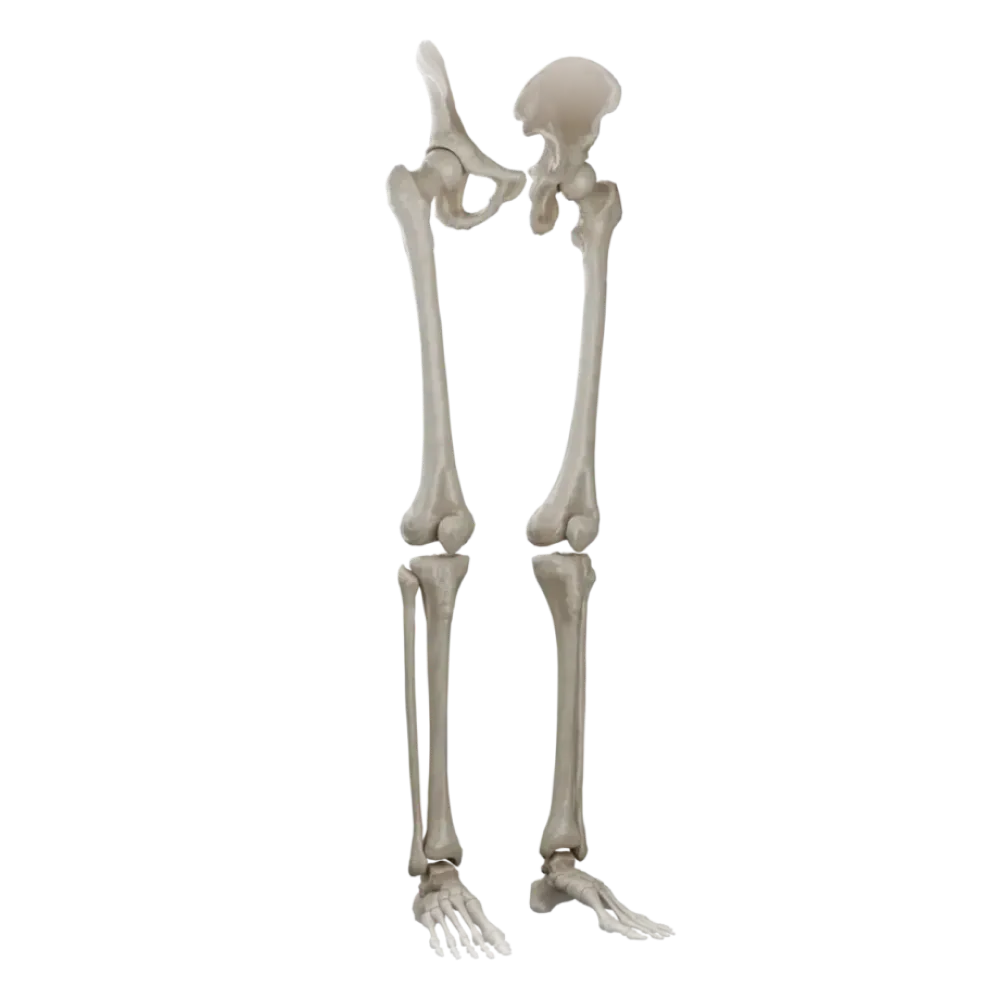

Test on the anatomy of the bones of the free lower limb.
Check the knowledge of anatomy of the bones of the free lower limb. The test covers the structure of the thigh, leg, and foot, and their articular surfaces.
1/20
bold
text
1. Which anatomical structure is located on the posterior surface of the femoral diaphysis?
-
Intertrochanteric line.
The linea aspera is located on the posterior surface of the femoral shaft and serves as an attachment site for muscles.
-
Patellar surface.
The linea aspera is located on the posterior surface of the femoral shaft and serves as an attachment site for muscles.
-
Linea aspera.
The linea aspera is located on the posterior surface of the femoral shaft and serves as an attachment site for muscles.
-
Intercondylar fossa.
The linea aspera is located on the posterior surface of the femoral shaft and serves as an attachment site for muscles.
-
I find it difficult to answer
The linea aspera is located on the posterior surface of the femoral shaft and serves as an attachment site for muscles.
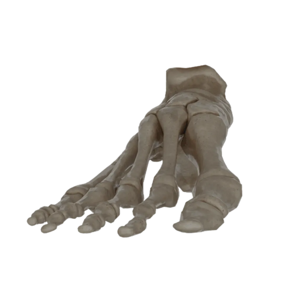
2. Which tarsal bone articulates with the leg bones, forming the ankle joint?
-
Calcaneus.
The talus forms a block for articulation with the leg bones, forming the ankle joint.
-
Talus.
The talus forms a block for articulation with the leg bones, forming the ankle joint.
-
Navicular bone.
The talus forms a block for articulation with the leg bones, forming the ankle joint.
-
Cuboid bone
The talus forms a block for articulation with the leg bones, forming the ankle joint.
-
I find it difficult to answer
The talus forms a block for articulation with the leg bones, forming the ankle joint.
3. Which structure is located on the anterior surface of the proximal epiphysis of the tibia and serves as the attachment point for the patellar ligament?
-
Medial condyle.
The tibial tuberosity is located on the anterior surface of the proximal epiphysis and serves as the attachment point for the patellar ligament.
-
Lateral condyle.
The tibial tuberosity is located on the anterior surface of the proximal epiphysis and serves as the attachment point for the patellar ligament.
-
Intercondylar eminence.
The tibial tuberosity is located on the anterior surface of the proximal epiphysis and serves as the attachment point for the patellar ligament.
-
Tibial tuberosity.
The tibial tuberosity is located on the anterior surface of the proximal epiphysis and serves as the attachment point for the patellar ligament.
-
I find it difficult to answer
The tibial tuberosity is located on the anterior surface of the proximal epiphysis and serves as the attachment point for the patellar ligament.
4. What structure separates the medial and lateral condyles on the posterior surface of the femur?
-
Intercondylar fossa.
The intercondylar fossa separates the medial and lateral condyles on the posterior surface of the femur.
-
Patellar surface.
The intercondylar fossa separates the medial and lateral condyles on the posterior surface of the femur.
-
Intertrochanteric crest.
The intercondylar fossa separates the medial and lateral condyles on the posterior surface of the femur.
-
Gluteal tuberosity.
The intercondylar fossa separates the medial and lateral condyles on the posterior surface of the femur.
-
I find it difficult to answer
The intercondylar fossa separates the medial and lateral condyles on the posterior surface of the femur.
5. Which bone does the cuboid bone articulate with distally?
-
With the navicular bone.
Distally, the cuboid bone articulates with the bases of the fourth and fifth metatarsal bones.
-
With the calcaneus.
Distally, the cuboid bone articulates with the bases of the fourth and fifth metatarsal bones.
-
With the first and second metatarsal bones.
Distally, the cuboid bone articulates with the bases of the fourth and fifth metatarsal bones.
-
With the fourth and fifth metatarsal bones.
Distally, the cuboid bone articulates with the bases of the fourth and fifth metatarsal bones.
-
I find it difficult to answer
Distally, the cuboid bone articulates with the bases of the fourth and fifth metatarsal bones.
6. Which part of the fibula participates in forming the ankle joint?
-
Head of the fibula.
The lateral malleolus is the distal end of the fibula and participates in forming the ankle joint.
-
Lateral malleolus.
The lateral malleolus is the distal end of the fibula and participates in forming the ankle joint.
-
Medial malleolus
The lateral malleolus is the distal end of the fibula and participates in forming the ankle joint.
-
Neck of the fibula.
The lateral malleolus is the distal end of the fibula and participates in forming the ankle joint.
-
I find it difficult to answer
The lateral malleolus is the distal end of the fibula and participates in forming the ankle joint.
7. Where is the groove for the tendon of the long fibular muscle located on the foot bones?
-
On the inferior surface of the cuboid bone.
The groove for the tendon of the long fibular muscle runs on the plantar surface of the cuboid bone.
-
On the superior surface of the talus.
The groove for the tendon of the long fibular muscle runs on the plantar surface of the cuboid bone.
-
On the medial surface of the navicular bone.
The groove for the tendon of the long fibular muscle runs on the plantar surface of the cuboid bone.
-
On the plantar surface of the calcaneus.
The groove for the tendon of the long fibular muscle runs on the plantar surface of the cuboid bone.
-
I find it difficult to answer
The groove for the tendon of the long fibular muscle runs on the plantar surface of the cuboid bone.
8. Which ligament attaches to the fovea of the femoral head?
-
Iliofemoral ligament.
The ligament of head of the femur attaches to the fovea of the head, containing nourishing vessels.
-
Ischiofemoral ligament.
The ligament of head of the femur attaches to the fovea of the head, containing nourishing vessels.
-
Ligament of head of femur.
The ligament of head of the femur attaches to the fovea of the head, containing nourishing vessels.
-
Pubofemoral ligament.
The ligament of head of the femur attaches to the fovea of the head, containing nourishing vessels.
-
I find it difficult to answer
The ligament of head of the femur attaches to the fovea of the head, containing nourishing vessels.
9. What structure is located on the calcaneus and serves as a support for the talus?
-
Sustentaculum tali.
The sustentaculum tali projects from the medial surface of the calcaneus and supports the talus.
-
Calcaneal tubercle.
The sustentaculum tali projects from the medial surface of the calcaneus and supports the talus.
-
Sulcus of the talus.
The sustentaculum tali projects from the medial surface of the calcaneus and supports the talus.
-
Lateral process of the calcaneal tubercle.
The sustentaculum tali projects from the medial surface of the calcaneus and supports the talus.
-
I find it difficult to answer
The sustentaculum tali projects from the medial surface of the calcaneus and supports the talus.
10. Which of the cuneiform bones articulates with the base of the first metatarsal bone?
-
Lateral cuneiform bone.
The medial cuneiform bone is the largest of the cuneiform bones and articulates with the base of the first metatarsal bone.
-
Intermediate cuneiform bone.
The medial cuneiform bone is the largest of the cuneiform bones and articulates with the base of the first metatarsal bone.
-
Cuboid bone
The medial cuneiform bone is the largest of the cuneiform bones and articulates with the base of the first metatarsal bone.
-
Medial cuneiform bone.
The medial cuneiform bone is the largest of the cuneiform bones and articulates with the base of the first metatarsal bone.
-
I find it difficult to answer
The medial cuneiform bone is the largest of the cuneiform bones and articulates with the base of the first metatarsal bone.
11. What structure is located on the posterior surface of the medial condyle of the tibia?
-
Groove for the tendon of the semimembranosus muscle.
On the posterior surface of the medial condyle of the tibia, there is a groove for the attachment of the tendon of the semimembranosus muscle.
-
Soleal line.
On the posterior surface of the medial condyle of the tibia, there is a groove for the attachment of the tendon of the semimembranosus muscle.
-
Tibial tuberosity.
On the posterior surface of the medial condyle of the tibia, there is a groove for the attachment of the tendon of the semimembranosus muscle.
-
Fibular articular facet.
On the posterior surface of the medial condyle of the tibia, there is a groove for the attachment of the tendon of the semimembranosus muscle.
-
I find it difficult to answer
On the posterior surface of the medial condyle of the tibia, there is a groove for the attachment of the tendon of the semimembranosus muscle.
12. On which end of the fibula is its head located?
-
Distal end.
The head of the fibula forms the proximal end of this bone.
-
Proximal end.
The head of the fibula forms the proximal end of this bone.
-
At the level of the mid-diaphysis.
The head of the fibula forms the proximal end of this bone.
-
On the lateral malleolus.
The head of the fibula forms the proximal end of this bone.
-
I find it difficult to answer
The head of the fibula forms the proximal end of this bone.
13. Which tarsal bone is located anterior to the talus and on the medial side of the foot?
-
Cuboid bone
The navicular bone is located on the medial margin of the foot, anterior to the talus and posterior to the cuneiform bones.
-
Calcaneus.
The navicular bone is located on the medial margin of the foot, anterior to the talus and posterior to the cuneiform bones.
-
Lateral cuneiform bone.
The navicular bone is located on the medial margin of the foot, anterior to the talus and posterior to the cuneiform bones.
-
Navicular bone.
The navicular bone is located on the medial margin of the foot, anterior to the talus and posterior to the cuneiform bones.
-
I find it difficult to answer
The navicular bone is located on the medial margin of the foot, anterior to the talus and posterior to the cuneiform bones.
14. Which anatomical structure is located on the posterior surface of the tibial diaphysis?
-
Soleal line.
The soleal line runs obliquely on the posterior surface of the tibial diaphysis from above downward and medially.
-
Anterior border
The soleal line runs obliquely on the posterior surface of the tibial diaphysis from above downward and medially.
-
Interosseous border.
The soleal line runs obliquely on the posterior surface of the tibial diaphysis from above downward and medially.
-
Medial surface
The soleal line runs obliquely on the posterior surface of the tibial diaphysis from above downward and medially.
-
I find it difficult to answer
The soleal line runs obliquely on the posterior surface of the tibial diaphysis from above downward and medially.
15. To which group of bones does the patella belong?
-
Spongy bones.
The patella is the largest sesamoid bone, embedded in the tendon of the quadriceps femoris muscle.
-
Flat bones
The patella is the largest sesamoid bone, embedded in the tendon of the quadriceps femoris muscle.
-
Sesamoid bones
The patella is the largest sesamoid bone, embedded in the tendon of the quadriceps femoris muscle.
-
Mixed bones.
The patella is the largest sesamoid bone, embedded in the tendon of the quadriceps femoris muscle.
-
I find it difficult to answer
The patella is the largest sesamoid bone, embedded in the tendon of the quadriceps femoris muscle.
16. How many phalanges does the first (big) toe have?
-
One.
The big toe (hallux) has only two phalanges: proximal and distal.
-
Two.
The big toe (hallux) has only two phalanges: proximal and distal.
-
Three.
The big toe (hallux) has only two phalanges: proximal and distal.
-
Four.
The big toe (hallux) has only two phalanges: proximal and distal.
-
I find it difficult to answer
The big toe (hallux) has only two phalanges: proximal and distal.
17. What is the thickening on the lateral side of the proximal epiphysis of the femur, to which muscles attach?
-
Lesser trochanter.
The greater trochanter represents a massive bony prominence on the lateral side of the proximal end of the femur.
-
Lateral epicondyle.
The greater trochanter represents a massive bony prominence on the lateral side of the proximal end of the femur.
-
Pectineal line
The greater trochanter represents a massive bony prominence on the lateral side of the proximal end of the femur.
-
Greater trochanter.
The greater trochanter represents a massive bony prominence on the lateral side of the proximal end of the femur.
-
I find it difficult to answer
The greater trochanter represents a massive bony prominence on the lateral side of the proximal end of the femur.
18. Which articular surface is located on the posterior side of the patella?
-
For the tibia.
The posterior (articular) surface of the patella articulates with the patellar surface of the femur.
-
For the femur.
The posterior (articular) surface of the patella articulates with the patellar surface of the femur.
-
For the fibula.
The posterior (articular) surface of the patella articulates with the patellar surface of the femur.
-
For the menisci.
The posterior (articular) surface of the patella articulates with the patellar surface of the femur.
-
I find it difficult to answer
The posterior (articular) surface of the patella articulates with the patellar surface of the femur.
19. Which foot bone forms the tarsal sinus (sinus tarsi) together with the talus?
-
Navicular bone.
The tarsal sinus (sinus tarsi) is formed by grooves on the opposite surfaces of the calcaneus and talus.
-
Calcaneus.
The tarsal sinus (sinus tarsi) is formed by grooves on the opposite surfaces of the calcaneus and talus.
-
Cuboid bone
The tarsal sinus (sinus tarsi) is formed by grooves on the opposite surfaces of the calcaneus and talus.
-
Medial cuneiform bone.
The tarsal sinus (sinus tarsi) is formed by grooves on the opposite surfaces of the calcaneus and talus.
-
I find it difficult to answer
The tarsal sinus (sinus tarsi) is formed by grooves on the opposite surfaces of the calcaneus and talus.
20. Where is the intertrochanteric crest located on the femur?
-
On the anterior surface between the trochanters.
The intertrochanteric crest (crista intertrochanterica) connects the greater and lesser trochanters on the posterior surface of the proximal epiphysis of the femur.
-
On the medial surface of the shaft.
The intertrochanteric crest (crista intertrochanterica) connects the greater and lesser trochanters on the posterior surface of the proximal epiphysis of the femur.
-
On the posterior surface between the trochanters.
The intertrochanteric crest (crista intertrochanterica) connects the greater and lesser trochanters on the posterior surface of the proximal epiphysis of the femur.
-
On the lateral surface of the condyle.
The intertrochanteric crest (crista intertrochanterica) connects the greater and lesser trochanters on the posterior surface of the proximal epiphysis of the femur.
-
I find it difficult to answer
The intertrochanteric crest (crista intertrochanterica) connects the greater and lesser trochanters on the posterior surface of the proximal epiphysis of the femur.
Retake this quiz?
Your current progress will be reset.