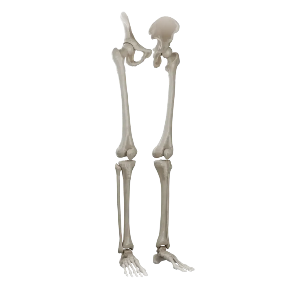
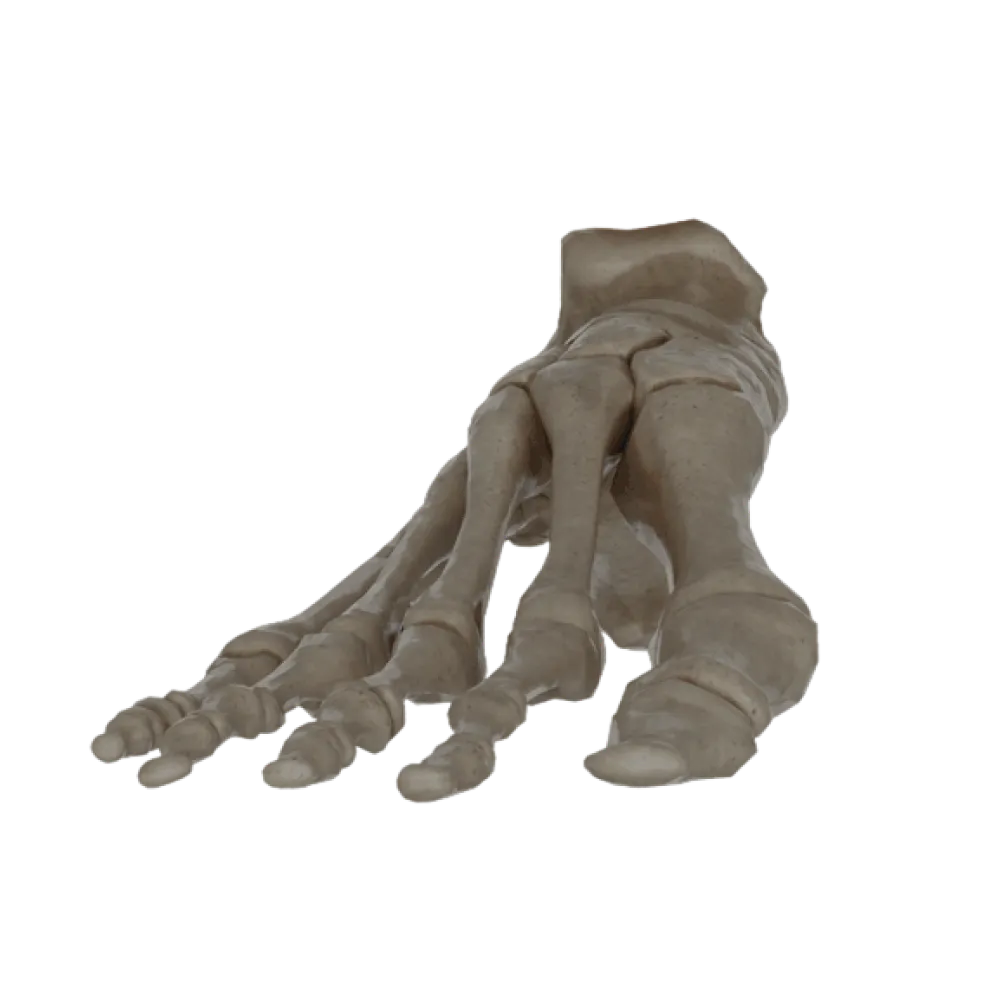

Foot bone anatomy test
Evaluate the knowledge of foot bone anatomy. The test assesses the topography of the tarsus, metatarsus, and phalanges, as well as the attachment sites of muscles and ligaments.
1/20
bold
text
1. Which tarsal bone articulates with the leg bones?
-
Calcaneus
The talus bone forms the ankle joint, articulating with the tibia and fibula.
-
Navicular
The talus bone forms the ankle joint, articulating with the tibia and fibula.
-
Talus
The talus bone forms the ankle joint, articulating with the tibia and fibula.
-
Cuboid
The talus bone forms the ankle joint, articulating with the tibia and fibula.
-
I find it difficult to answer
The talus bone forms the ankle joint, articulating with the tibia and fibula.
2. On which bone is the sustentaculum tali located?
-
On the inferior surface of the talus
The sustentaculum tali is a process on the medial surface of the calcaneus, supporting the talus.
-
On the medial surface of the calcaneus
The sustentaculum tali is a process on the medial surface of the calcaneus, supporting the talus.
-
On the lateral surface of the calcaneus.
The sustentaculum tali is a process on the medial surface of the calcaneus, supporting the talus.
-
On the medial cuneiform bone
The sustentaculum tali is a process on the medial surface of the calcaneus, supporting the talus.
-
I find it difficult to answer
The sustentaculum tali is a process on the medial surface of the calcaneus, supporting the talus.
3. Which muscle attaches to the tuberosity of the navicular bone?
-
Tibialis anterior muscle.
The tuberosity of the navicular bone is the primary attachment site of the tibialis posterior tendon.
-
Fibularis longus muscle.
The tuberosity of the navicular bone is the primary attachment site of the tibialis posterior tendon.
-
Fibularis brevis muscle.
The tuberosity of the navicular bone is the primary attachment site of the tibialis posterior tendon.
-
Tibialis posterior muscle.
The tuberosity of the navicular bone is the primary attachment site of the tibialis posterior tendon.
-
I find it difficult to answer
The tuberosity of the navicular bone is the primary attachment site of the tibialis posterior tendon.
4. On the plantar surface of which bone is the groove for the fibularis longus tendon located?
-
Cuboid
The groove for the tendon of the fibularis longus is located on the plantar surface of the cuboid bone.
-
Calcaneus
The groove for the tendon of the fibularis longus is located on the plantar surface of the cuboid bone.
-
Navicular
The groove for the tendon of the fibularis longus is located on the plantar surface of the cuboid bone.
-
Medial cuneiform
The groove for the tendon of the fibularis longus is located on the plantar surface of the cuboid bone.
-
I find it difficult to answer
The groove for the tendon of the fibularis longus is located on the plantar surface of the cuboid bone.
5. Which metatarsal bone does the medial cuneiform bone articulate with?
-
Second
The medial cuneiform bone (os cuneiforme mediale) is the largest and articulates with the base of the first metatarsal bone.
-
Third
The medial cuneiform bone (os cuneiforme mediale) is the largest and articulates with the base of the first metatarsal bone.
-
First
The medial cuneiform bone (os cuneiforme mediale) is the largest and articulates with the base of the first metatarsal bone.
-
Fourth and fifth
The medial cuneiform bone (os cuneiforme mediale) is the largest and articulates with the base of the first metatarsal bone.
-
I find it difficult to answer
The medial cuneiform bone (os cuneiforme mediale) is the largest and articulates with the base of the first metatarsal bone.
6. What structure is located on the lateral side of the base of the fifth metatarsal bone?
-
Tuberosity of the fifth metatarsal bone
The tuberosity of the fifth metatarsal bone (tuberositas ossis metatarsi quinti) serves as the attachment site for the fibularis brevis muscle.
-
Sustentaculum tali
The tuberosity of the fifth metatarsal bone (tuberositas ossis metatarsi quinti) serves as the attachment site for the fibularis brevis muscle.
-
Trochlea of talus
The tuberosity of the fifth metatarsal bone (tuberositas ossis metatarsi quinti) serves as the attachment site for the fibularis brevis muscle.
-
Medial malleolus
The tuberosity of the fifth metatarsal bone (tuberositas ossis metatarsi quinti) serves as the attachment site for the fibularis brevis muscle.
-
I find it difficult to answer
The tuberosity of the fifth metatarsal bone (tuberositas ossis metatarsi quinti) serves as the attachment site for the fibularis brevis muscle.
7. How many phalanges does the first toe (hallux) have?
-
One.
The big toe, like the thumb, has only two phalanges: proximal and distal.
-
Three.
The big toe, like the thumb, has only two phalanges: proximal and distal.
-
Four.
The big toe, like the thumb, has only two phalanges: proximal and distal.
-
Two.
The big toe, like the thumb, has only two phalanges: proximal and distal.
-
I find it difficult to answer
The big toe, like the thumb, has only two phalanges: proximal and distal.
8. Between which bones does the tarsal sinus (sinus tarsi) form?
-
Between the calcaneus and cuboid bones
The tarsal sinus is formed by grooves in the talus and calcaneus bones and contains the interosseous talocalcaneal ligament.
-
Between the talus and calcaneus bones
The tarsal sinus is formed by grooves in the talus and calcaneus bones and contains the interosseous talocalcaneal ligament.
-
Between the talus and navicular bones
The tarsal sinus is formed by grooves in the talus and calcaneus bones and contains the interosseous talocalcaneal ligament.
-
Between the cuneiform and metatarsal bones
The tarsal sinus is formed by grooves in the talus and calcaneus bones and contains the interosseous talocalcaneal ligament.
-
I find it difficult to answer
The tarsal sinus is formed by grooves in the talus and calcaneus bones and contains the interosseous talocalcaneal ligament.
9. Which muscle's tendon passes through the groove on the posterior process of the talus?
-
Flexor digitorum longus
The groove for the tendon of the flexor hallucis longus muscle passes between the medial and lateral tubercles of the posterior process of the talus.
-
Tibialis posterior muscle
The groove for the tendon of the flexor hallucis longus muscle passes between the medial and lateral tubercles of the posterior process of the talus.
-
Flexor hallucis longus
The groove for the tendon of the flexor hallucis longus muscle passes between the medial and lateral tubercles of the posterior process of the talus.
-
Extensor hallucis longus
The groove for the tendon of the flexor hallucis longus muscle passes between the medial and lateral tubercles of the posterior process of the talus.
-
I find it difficult to answer
The groove for the tendon of the flexor hallucis longus muscle passes between the medial and lateral tubercles of the posterior process of the talus.
10. Which structure does the calcaneal (Achilles) tendon attach to?
-
Posterior process of talus
The calcaneal tendon (tendo calcaneus) attaches to the calcaneal tuberosity (tuber calcanei).
-
Tuberosity of navicular bone
The calcaneal tendon (tendo calcaneus) attaches to the calcaneal tuberosity (tuber calcanei).
-
Sustentaculum tali
The calcaneal tendon (tendo calcaneus) attaches to the calcaneal tuberosity (tuber calcanei).
-
Calcaneal tuberosity
The calcaneal tendon (tendo calcaneus) attaches to the calcaneal tuberosity (tuber calcanei).
-
I find it difficult to answer
The calcaneal tendon (tendo calcaneus) attaches to the calcaneal tuberosity (tuber calcanei).
11. Which bones form the transverse tarsal joint (Chopart's joint)?
-
Talonavicular and calcaneocuboid joints
The Chopart joint (articulatio tarsi transversa) functionally unites the talonavicular and calcaneocuboid joints.
-
Cuneonavicular joint
The Chopart joint (articulatio tarsi transversa) functionally unites the talonavicular and calcaneocuboid joints.
-
Tarsometatarsal joints
The Chopart joint (articulatio tarsi transversa) functionally unites the talonavicular and calcaneocuboid joints.
-
Subtalar joint
The Chopart joint (articulatio tarsi transversa) functionally unites the talonavicular and calcaneocuboid joints.
-
I find it difficult to answer
The Chopart joint (articulatio tarsi transversa) functionally unites the talonavicular and calcaneocuboid joints.
12. Which bone is the keystone (highest point) of the medial longitudinal arch of the foot?
-
Calcaneus
The talus bone transmits body weight to the foot arches and is the apex of the medial longitudinal arch.
-
Navicular
The talus bone transmits body weight to the foot arches and is the apex of the medial longitudinal arch.
-
Talus
The talus bone transmits body weight to the foot arches and is the apex of the medial longitudinal arch.
-
Medial cuneiform
The talus bone transmits body weight to the foot arches and is the apex of the medial longitudinal arch.
-
I find it difficult to answer
The talus bone transmits body weight to the foot arches and is the apex of the medial longitudinal arch.
13. Which muscle's tendon passes directly beneath the sustentaculum tali?
-
Flexor digitorum longus
The tendon of the flexor hallucis longus (m. flexor hallucis longus) passes in the groove under the sustentaculum tali of the calcaneus.
-
Flexor hallucis longus
The tendon of the flexor hallucis longus (m. flexor hallucis longus) passes in the groove under the sustentaculum tali of the calcaneus.
-
Tibialis posterior muscle
The tendon of the flexor hallucis longus (m. flexor hallucis longus) passes in the groove under the sustentaculum tali of the calcaneus.
-
Tibialis anterior muscle
The tendon of the flexor hallucis longus (m. flexor hallucis longus) passes in the groove under the sustentaculum tali of the calcaneus.
-
I find it difficult to answer
The tendon of the flexor hallucis longus (m. flexor hallucis longus) passes in the groove under the sustentaculum tali of the calcaneus.
14. To which metatarsal bone's base does the tendon of the tibialis anterior muscle attach?
-
Second
The tendon of the tibialis anterior muscle attaches to the medial cuneiform bone and the base of the first metatarsal bone.
-
Third
The tendon of the tibialis anterior muscle attaches to the medial cuneiform bone and the base of the first metatarsal bone.
-
Fifth
The tendon of the tibialis anterior muscle attaches to the medial cuneiform bone and the base of the first metatarsal bone.
-
First
The tendon of the tibialis anterior muscle attaches to the medial cuneiform bone and the base of the first metatarsal bone.
-
I find it difficult to answer
The tendon of the tibialis anterior muscle attaches to the medial cuneiform bone and the base of the first metatarsal bone.
15. Which bones articulate distally with the cuboid bone?
-
Fourth and fifth metatarsal bones
The cuboid bone distally forms joints with the bases of the fourth and fifth metatarsal bones.
-
Calcaneus and navicular
The cuboid bone distally forms joints with the bases of the fourth and fifth metatarsal bones.
-
Talus and calcaneus
The cuboid bone distally forms joints with the bases of the fourth and fifth metatarsal bones.
-
First and second metatarsal bones
The cuboid bone distally forms joints with the bases of the fourth and fifth metatarsal bones.
-
I find it difficult to answer
The cuboid bone distally forms joints with the bases of the fourth and fifth metatarsal bones.
16. Which structure articulates with the anterior articular surface of the calcaneus?
-
With the navicular bone.
The anterior cuboid articular surface of the calcaneus (facies articularis cuboidea) articulates with the cuboid bone.
-
With the talus
The anterior cuboid articular surface of the calcaneus (facies articularis cuboidea) articulates with the cuboid bone.
-
With the cuboid bone
The anterior cuboid articular surface of the calcaneus (facies articularis cuboidea) articulates with the cuboid bone.
-
With the lateral cuneiform bone
The anterior cuboid articular surface of the calcaneus (facies articularis cuboidea) articulates with the cuboid bone.
-
I find it difficult to answer
The anterior cuboid articular surface of the calcaneus (facies articularis cuboidea) articulates with the cuboid bone.
17. Where are the sesamoid bones of the foot most commonly located?
-
In the region of the base of the fifth metatarsal bone
Two large sesamoid bones (medial and lateral) are consistently present in the flexor tendons beneath the head of the first metatarsal bone.
-
In the region of the head of the first metatarsal bone
Two large sesamoid bones (medial and lateral) are consistently present in the flexor tendons beneath the head of the first metatarsal bone.
-
Within the thickness of the Achilles tendon
Two large sesamoid bones (medial and lateral) are consistently present in the flexor tendons beneath the head of the first metatarsal bone.
-
In the region of the tuberosity of the navicular bone
Two large sesamoid bones (medial and lateral) are consistently present in the flexor tendons beneath the head of the first metatarsal bone.
-
I find it difficult to answer
Two large sesamoid bones (medial and lateral) are consistently present in the flexor tendons beneath the head of the first metatarsal bone.
18. The Lisfranc ligament (lig. cuneometatarsale interosseum mediale) connects:
-
the navicular and cuboid bones.
This ligament is the key of the Lisfranc joint and extends from the medial cuneiform bone to the base of the II metatarsal bone.
-
the cuboid and V metatarsal bones.
This ligament is the key of the Lisfranc joint and extends from the medial cuneiform bone to the base of the II metatarsal bone.
-
the talar and navicular bones.
This ligament is the key of the Lisfranc joint and extends from the medial cuneiform bone to the base of the II metatarsal bone.
-
the medial cuneiform bone and the base of the II metatarsal bone;
This ligament is the key of the Lisfranc joint and extends from the medial cuneiform bone to the base of the II metatarsal bone.
-
I find it difficult to answer
This ligament is the key of the Lisfranc joint and extends from the medial cuneiform bone to the base of the II metatarsal bone.
19. On which bone is the fibular trochlea located?
-
On the lateral surface of the calcaneus.
The fibular trochlea is located on the lateral surface of the calcaneus, dividing the tendons of the fibular muscles.
-
On the medial surface of the talus.
The fibular trochlea is located on the lateral surface of the calcaneus, dividing the tendons of the fibular muscles.
-
On the lateral surface of the cuboid bone.
The fibular trochlea is located on the lateral surface of the calcaneus, dividing the tendons of the fibular muscles.
-
On the lateral malleolus.
The fibular trochlea is located on the lateral surface of the calcaneus, dividing the tendons of the fibular muscles.
-
I find it difficult to answer
The fibular trochlea is located on the lateral surface of the calcaneus, dividing the tendons of the fibular muscles.
20. What articular surfaces are located on the underside of the talus?
-
the navicular and cuboid articular surfaces.
On the inferior surface of the talus are three calcaneal articular surfaces (anterior, middle, posterior) for articulation with the calcaneus.
-
the anterior, middle, and posterior calcaneal articular surfaces.
On the inferior surface of the talus are three calcaneal articular surfaces (anterior, middle, posterior) for articulation with the calcaneus.
-
the medial and lateral malleolar surfaces.
On the inferior surface of the talus are three calcaneal articular surfaces (anterior, middle, posterior) for articulation with the calcaneus.
-
the superior and inferior cuneiform surfaces.
On the inferior surface of the talus are three calcaneal articular surfaces (anterior, middle, posterior) for articulation with the calcaneus.
-
I find it difficult to answer
On the inferior surface of the talus are three calcaneal articular surfaces (anterior, middle, posterior) for articulation with the calcaneus.
Retake this quiz?
Your current progress will be reset.