1/20
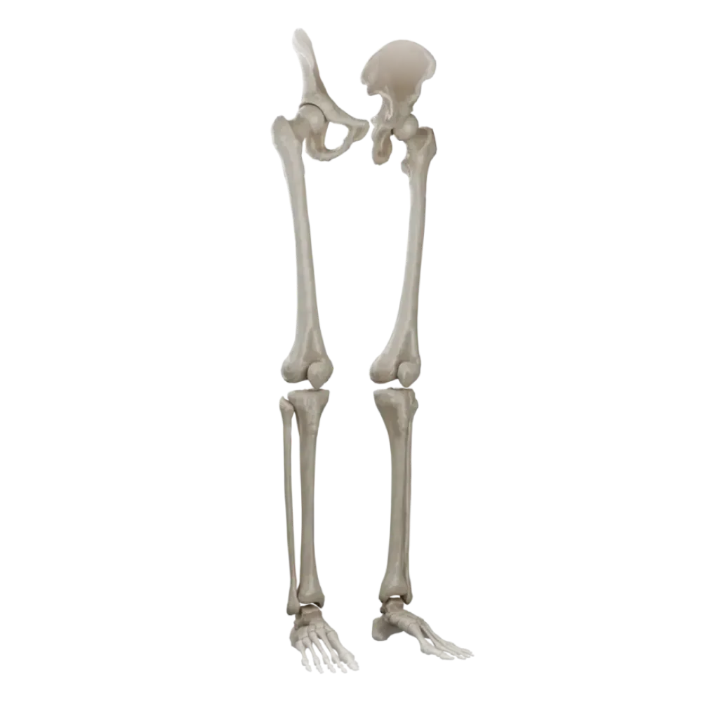

Test on the anatomy of the bones of the lower limb.
Evaluate the knowledge of osteology of the lower limb. The test strictly evaluates the understanding of topography, articular surfaces, and bony landmarks.
1. Which bones form the acetabulum?
-
Ilium, ischium, and pubis.
The acetabulum is formed by the bodies of three pelvic bones: ilium, pubis, and ischium, which fuse at the area of the Y-shaped cartilage.
-
Ilium and sacrum.
The acetabulum is formed by the bodies of three pelvic bones: ilium, pubis, and ischium, which fuse at the area of the Y-shaped cartilage.
-
Ischium and pubis.
The acetabulum is formed by the bodies of three pelvic bones: ilium, pubis, and ischium, which fuse at the area of the Y-shaped cartilage.
-
Ilium, pubis, and sacrum.
The acetabulum is formed by the bodies of three pelvic bones: ilium, pubis, and ischium, which fuse at the area of the Y-shaped cartilage.
-
I find it difficult to answer
The acetabulum is formed by the bodies of three pelvic bones: ilium, pubis, and ischium, which fuse at the area of the Y-shaped cartilage.
2. On which surface of the femur is the linea aspera located?
-
Anterior.
The linea aspera is located on the posterior surface of the femoral shaft and serves as an attachment site for muscles.
-
Medial.
The linea aspera is located on the posterior surface of the femoral shaft and serves as an attachment site for muscles.
-
Posterior.
The linea aspera is located on the posterior surface of the femoral shaft and serves as an attachment site for muscles.
-
Lateral.
The linea aspera is located on the posterior surface of the femoral shaft and serves as an attachment site for muscles.
-
I find it difficult to answer
The linea aspera is located on the posterior surface of the femoral shaft and serves as an attachment site for muscles.
3. To which anatomical structure of the tibia does the patellar ligament attach?
-
Tibial tuberosity.
The tibial tuberosity is the site of attachment for the patellar ligament.
-
Medial condyle.
The tibial tuberosity is the site of attachment for the patellar ligament.
-
Intercondylar eminence.
The tibial tuberosity is the site of attachment for the patellar ligament.
-
Lateral condyle.
The tibial tuberosity is the site of attachment for the patellar ligament.
-
I find it difficult to answer
The tibial tuberosity is the site of attachment for the patellar ligament.
4. Which bones bound the obturator foramen of the pelvic bone?
-
Ilium and pubis.
The obturator foramen is bounded by the branches of the pubis and ischium.
-
Ischium and ilium.
The obturator foramen is bounded by the branches of the pubis and ischium.
-
Only pubis.
The obturator foramen is bounded by the branches of the pubis and ischium.
-
Pubis and ischium.
The obturator foramen is bounded by the branches of the pubis and ischium.
-
I find it difficult to answer
The obturator foramen is bounded by the branches of the pubis and ischium.
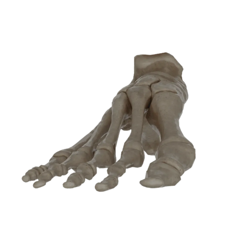
5. Which tarsal bones articulate proximally with the cuboid bone?
-
Talus.
Proximally, the cuboid bone articulates with the cuboid articular surface of the calcaneus.
-
Navicular bone.
Proximally, the cuboid bone articulates with the cuboid articular surface of the calcaneus.
-
Medial cuneiform bone.
Proximally, the cuboid bone articulates with the cuboid articular surface of the calcaneus.
-
Calcaneus.
Proximally, the cuboid bone articulates with the cuboid articular surface of the calcaneus.
-
I find it difficult to answer
Proximally, the cuboid bone articulates with the cuboid articular surface of the calcaneus.
6. Which structure separates the greater and lesser sciatic notches?
-
Ischial tuberosity.
The ischial spine, located on the posterior edge of the ischium, projects between the greater and lesser sciatic notches.
-
Pubic tubercle.
The ischial spine, located on the posterior edge of the ischium, projects between the greater and lesser sciatic notches.
-
Ischial spine.
The ischial spine, located on the posterior edge of the ischium, projects between the greater and lesser sciatic notches.
-
Iliac crest.
The ischial spine, located on the posterior edge of the ischium, projects between the greater and lesser sciatic notches.
-
I find it difficult to answer
The ischial spine, located on the posterior edge of the ischium, projects between the greater and lesser sciatic notches.
7. Where is the lateral malleolus located?
-
On the distal epiphysis of the tibia.
The lateral malleolus is the thickened distal end of the fibula.
-
On the proximal epiphysis of the fibula.
The lateral malleolus is the thickened distal end of the fibula.
-
On the distal epiphysis of the fibula.
The lateral malleolus is the thickened distal end of the fibula.
-
On the talus bone.
The lateral malleolus is the thickened distal end of the fibula.
-
I find it difficult to answer
The lateral malleolus is the thickened distal end of the fibula.
8. On which surface of the patella is the articular surface located?
-
Anterior.
The articular surface of the patella, covered with hyaline cartilage, faces posteriorly, towards the patellar surface on the femur.
-
Superior.
The articular surface of the patella, covered with hyaline cartilage, faces posteriorly, towards the patellar surface on the femur.
-
Inferior.
The articular surface of the patella, covered with hyaline cartilage, faces posteriorly, towards the patellar surface on the femur.
-
Posterior.
The articular surface of the patella, covered with hyaline cartilage, faces posteriorly, towards the patellar surface on the femur.
-
I find it difficult to answer
The articular surface of the patella, covered with hyaline cartilage, faces posteriorly, towards the patellar surface on the femur.
9. Which bone does the navicular bone of the foot articulate proximally with?
-
Head of the talus.
The proximal concave articular surface of the navicular bone articulates with the head of the talus, forming the talonavicular joint.
-
Calcaneus.
The proximal concave articular surface of the navicular bone articulates with the head of the talus, forming the talonavicular joint.
-
Cuboid bone
The proximal concave articular surface of the navicular bone articulates with the head of the talus, forming the talonavicular joint.
-
Medial cuneiform bone.
The proximal concave articular surface of the navicular bone articulates with the head of the talus, forming the talonavicular joint.
-
I find it difficult to answer
The proximal concave articular surface of the navicular bone articulates with the head of the talus, forming the talonavicular joint.
10. What formation separates the medial and lateral condyles of the tibia at its proximal end?
-
Intercondylar fossa.
The intercondylar eminence is located on the superior articular surface, between the condyles of the tibia.
-
Soleal line.
The intercondylar eminence is located on the superior articular surface, between the condyles of the tibia.
-
Tibial tuberosity.
The intercondylar eminence is located on the superior articular surface, between the condyles of the tibia.
-
Intercondylar eminence.
The intercondylar eminence is located on the superior articular surface, between the condyles of the tibia.
-
I find it difficult to answer
The intercondylar eminence is located on the superior articular surface, between the condyles of the tibia.
11. On which tarsal bone is the sustentaculum tali located?
-
Talus.
The sustentaculum tali is a process of the calcaneus, projecting medially and supporting the head of the talus.
-
Cuboid bone
The sustentaculum tali is a process of the calcaneus, projecting medially and supporting the head of the talus.
-
Navicular bone.
The sustentaculum tali is a process of the calcaneus, projecting medially and supporting the head of the talus.
-
Calcaneus.
The sustentaculum tali is a process of the calcaneus, projecting medially and supporting the head of the talus.
-
I find it difficult to answer
The sustentaculum tali is a process of the calcaneus, projecting medially and supporting the head of the talus.
12. Where is the intertrochanteric crest located?
-
On the posterior surface of the proximal end of the femur.
The intertrochanteric crest connects the greater and lesser trochanters on the posterior surface of the femur.
-
On the anterior surface of the proximal end of the femur.
The intertrochanteric crest connects the greater and lesser trochanters on the posterior surface of the femur.
-
On the distal epiphysis of the femur.
The intertrochanteric crest connects the greater and lesser trochanters on the posterior surface of the femur.
-
Between the condyles of the tibia.
The intertrochanteric crest connects the greater and lesser trochanters on the posterior surface of the femur.
-
I find it difficult to answer
The intertrochanteric crest connects the greater and lesser trochanters on the posterior surface of the femur.
13. Which bone has a medial malleolus?
-
Fibula
The medial malleolus is the medial projection of the distal epiphysis of the tibia.
-
Talus.
The medial malleolus is the medial projection of the distal epiphysis of the tibia.
-
Tibia
The medial malleolus is the medial projection of the distal epiphysis of the tibia.
-
Calcaneus.
The medial malleolus is the medial projection of the distal epiphysis of the tibia.
-
I find it difficult to answer
The medial malleolus is the medial projection of the distal epiphysis of the tibia.
14. Where is the fovea capitis of the femoral head located?
-
On the neck of the femur.
The fovea capitis is located slightly below and posterior to the center of the femoral head; to it attaches the ligament of the head of the femur.
-
In the intercondylar fossa.
The fovea capitis is located slightly below and posterior to the center of the femoral head; to it attaches the ligament of the head of the femur.
-
On the medial surface of the femoral head.
The fovea capitis is located slightly below and posterior to the center of the femoral head; to it attaches the ligament of the head of the femur.
-
On the acetabulum.
The fovea capitis is located slightly below and posterior to the center of the femoral head; to it attaches the ligament of the head of the femur.
-
I find it difficult to answer
The fovea capitis is located slightly below and posterior to the center of the femoral head; to it attaches the ligament of the head of the femur.
15. Which tarsal bones do the three cuneiform bones articulate distally with?
-
With the first, second, and third metatarsal bones.
The medial, intermediate, and lateral cuneiform bones articulate with the bases of the first, second, and third metatarsal bones, respectively.
-
With the third, fourth, and fifth metatarsal bones.
The medial, intermediate, and lateral cuneiform bones articulate with the bases of the first, second, and third metatarsal bones, respectively.
-
With the navicular bone.
The medial, intermediate, and lateral cuneiform bones articulate with the bases of the first, second, and third metatarsal bones, respectively.
-
With the cuboid bone
The medial, intermediate, and lateral cuneiform bones articulate with the bases of the first, second, and third metatarsal bones, respectively.
-
I find it difficult to answer
The medial, intermediate, and lateral cuneiform bones articulate with the bases of the first, second, and third metatarsal bones, respectively.
16. On which bone is the iliac crest located?
-
Ischium
The iliac crest is the superior thickened border of the wing of the ilium.
-
Ilium
The iliac crest is the superior thickened border of the wing of the ilium.
-
Sacrum.
The iliac crest is the superior thickened border of the wing of the ilium.
-
Pubis
The iliac crest is the superior thickened border of the wing of the ilium.
-
I find it difficult to answer
The iliac crest is the superior thickened border of the wing of the ilium.
17. Which part of the fibula has an articular surface for articulating with the tibia proximally?
-
Lateral malleolus.
The head of the fibula carries an articular surface for articulation with the lateral condyle of the tibia.
-
Head of the fibula.
The head of the fibula carries an articular surface for articulation with the lateral condyle of the tibia.
-
Neck of the fibula.
The head of the fibula carries an articular surface for articulation with the lateral condyle of the tibia.
-
Body of the fibula.
The head of the fibula carries an articular surface for articulation with the lateral condyle of the tibia.
-
I find it difficult to answer
The head of the fibula carries an articular surface for articulation with the lateral condyle of the tibia.
18. What forms the inferior boundary of the greater pelvis and the superior boundary of the lesser pelvis?
-
Linea aspera.
The terminal line separates the greater and lesser pelvis, passing over the promontory of the sacrum and arcuate lines of the ilium.
-
Linea terminalis.
The terminal line separates the greater and lesser pelvis, passing over the promontory of the sacrum and arcuate lines of the ilium.
-
Pectineal line
The terminal line separates the greater and lesser pelvis, passing over the promontory of the sacrum and arcuate lines of the ilium.
-
Intertrochanteric line
The terminal line separates the greater and lesser pelvis, passing over the promontory of the sacrum and arcuate lines of the ilium.
-
I find it difficult to answer
The terminal line separates the greater and lesser pelvis, passing over the promontory of the sacrum and arcuate lines of the ilium.
19. How many phalanges does the first toe (hallux) have?
-
Three.
The big toe (hallux), like the thumb, has only two phalanges: proximal and distal.
-
Two.
The big toe (hallux), like the thumb, has only two phalanges: proximal and distal.
-
One.
The big toe (hallux), like the thumb, has only two phalanges: proximal and distal.
-
Four.
The big toe (hallux), like the thumb, has only two phalanges: proximal and distal.
-
I find it difficult to answer
The big toe (hallux), like the thumb, has only two phalanges: proximal and distal.
20. Which muscle attaches to the lesser trochanter of the femur?
-
Gluteus maximus muscle.
The lesser trochanter serves as the attachment site for the powerful iliopsoas muscle, which flexes the thigh.
-
Iliopsoas muscle
The lesser trochanter serves as the attachment site for the powerful iliopsoas muscle, which flexes the thigh.
-
Biceps femoris muscle.
The lesser trochanter serves as the attachment site for the powerful iliopsoas muscle, which flexes the thigh.
-
Quadriceps femoris muscle.
The lesser trochanter serves as the attachment site for the powerful iliopsoas muscle, which flexes the thigh.
-
I find it difficult to answer
The lesser trochanter serves as the attachment site for the powerful iliopsoas muscle, which flexes the thigh.
Retake this quiz?
Your current progress will be reset.
Bones of the lower limb.
Bones of the free lower limb.
0/20