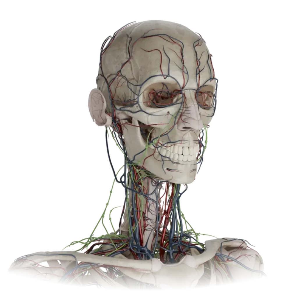
Anatomy test of the external carotid artery
Evaluate the knowledge of the anatomy of the external carotid artery. The test examines syn- topia, branching, anastomoses, and areas of blood supply at the university level.
1/20
bold
text
1. At which anatomical landmark does the bifurcation of the common carotid artery most frequently occur into the external and internal carotid arteries?
-
Hyoid bone.
The bifurcation of the common carotid artery in the carotid triangle is usually projected at the level of the upper border of the thyroid cartilage (corresponding to vertebra C4).
-
Angle of the mandible.
The bifurcation of the common carotid artery in the carotid triangle is usually projected at the level of the upper border of the thyroid cartilage (corresponding to vertebra C4).
-
The upper border of the thyroid cartilage.
The bifurcation of the common carotid artery in the carotid triangle is usually projected at the level of the upper border of the thyroid cartilage (corresponding to vertebra C4).
-
Cricoid cartilage.
The bifurcation of the common carotid artery in the carotid triangle is usually projected at the level of the upper border of the thyroid cartilage (corresponding to vertebra C4).
-
I find it difficult to answer
The bifurcation of the common carotid artery in the carotid triangle is usually projected at the level of the upper border of the thyroid cartilage (corresponding to vertebra C4).
2. Which of the listed arteries does NOT belong to the anterior group of branches of the external carotid artery?
-
Superior thyroid artery.
The ascending pharyngeal artery is the only medial branch of the external carotid artery. The anterior group includes the superior thyroid, lingual, and facial arteries.
-
Lingual artery.
The ascending pharyngeal artery is the only medial branch of the external carotid artery. The anterior group includes the superior thyroid, lingual, and facial arteries.
-
Facial artery.
The ascending pharyngeal artery is the only medial branch of the external carotid artery. The anterior group includes the superior thyroid, lingual, and facial arteries.
-
Ascending pharyngeal artery.
The ascending pharyngeal artery is the only medial branch of the external carotid artery. The anterior group includes the superior thyroid, lingual, and facial arteries.
-
I find it difficult to answer
The ascending pharyngeal artery is the only medial branch of the external carotid artery. The anterior group includes the superior thyroid, lingual, and facial arteries.
3. What is the typical syntopic relationship between the external (ECA) and internal (ICA) carotid arteries immediately after bifurcation?
-
The ECA is located anteriorly and medially to the ICA.
At the site of bifurcation, the external carotid artery is usually located somewhat anterior and medial relative to the internal carotid artery.
-
The ECA is located posteriorly and laterally to the ICA.
At the site of bifurcation, the external carotid artery is usually located somewhat anterior and medial relative to the internal carotid artery.
-
The ECA is strictly lateral to the ICA.
At the site of bifurcation, the external carotid artery is usually located somewhat anterior and medial relative to the internal carotid artery.
-
The ECA is strictly medial to the ICA.
At the site of bifurcation, the external carotid artery is usually located somewhat anterior and medial relative to the internal carotid artery.
-
I find it difficult to answer
At the site of bifurcation, the external carotid artery is usually located somewhat anterior and medial relative to the internal carotid artery.
4. In Pirogov's triangle (lingual triangle), the lingual artery is situated:
-
Superficial to the hyoglossus muscle (m. hyoglossus).
The lingual artery passes in Pirogov's triangle, lying deeper than the hypoglossal nerve and lingual vein. the hyoglossus, which separates it from the lingual vein and hypoglossal nerve.
-
Deeper than the hyoglossus muscle, on the middle pharyngeal constrictors.
The lingual artery passes in Pirogov's triangle, lying deeper than the hypoglossal nerve and lingual vein. the hyoglossus, which separates it from the lingual vein and hypoglossal nerve.
-
Between the mylohyoid and digastric muscles.
The lingual artery passes in Pirogov's triangle, lying deeper than the hypoglossal nerve and lingual vein. the hyoglossus, which separates it from the lingual vein and hypoglossal nerve.
-
Subcutaneously in the submandibular triangle.
The lingual artery passes in Pirogov's triangle, lying deeper than the hypoglossal nerve and lingual vein. the hyoglossus, which separates it from the lingual vein and hypoglossal nerve.
-
I find it difficult to answer
The lingual artery passes in Pirogov's triangle, lying deeper than the hypoglossal nerve and lingual vein. the hyoglossus, which separates it from the lingual vein and hypoglossal nerve.
5. Into which terminal branches does the external carotid artery divide within the parotid gland?
-
Superficial temporal artery and transverse facial artery.
At the level of the neck of the mandible in the parenchyma of the parotid gland, the external carotid artery divides into its terminal branches. maxillaris and a. superficialis temporalis.
-
Facial artery and lingual artery.
At the level of the neck of the mandible in the parenchyma of the parotid gland, the external carotid artery divides into its terminal branches. maxillaris and a. superficialis temporalis.
-
Posterior auricular artery and occipital artery.
At the level of the neck of the mandible in the parenchyma of the parotid gland, the external carotid artery divides into its terminal branches. maxillaris and a. superficialis temporalis.
-
Maxillary artery and superficial temporal artery.
At the level of the neck of the mandible in the parenchyma of the parotid gland, the external carotid artery divides into its terminal branches. maxillaris and a. superficialis temporalis.
-
I find it difficult to answer
At the level of the neck of the mandible in the parenchyma of the parotid gland, the external carotid artery divides into its terminal branches. maxillaris and a. superficialis temporalis.
6. Which artery is the terminal branch of the facial artery in the region of the medial angle of the eye?
-
Infraorbital artery.
The angular artery (a. angularis) is the terminal part of the facial artery, anastomosing with the dorsal nasal artery (a branch of the ophthalmic artery from the internal carotid system).
-
Transverse facial artery.
The angular artery (a. angularis) is the terminal part of the facial artery, anastomosing with the dorsal nasal artery (a branch of the ophthalmic artery from the internal carotid system).
-
Dorsal nasal artery.
The angular artery (a. angularis) is the terminal part of the facial artery, anastomosing with the dorsal nasal artery (a branch of the ophthalmic artery from the internal carotid system).
-
Angular artery.
The angular artery (a. angularis) is the terminal part of the facial artery, anastomosing with the dorsal nasal artery (a branch of the ophthalmic artery from the internal carotid system).
-
I find it difficult to answer
The angular artery (a. angularis) is the terminal part of the facial artery, anastomosing with the dorsal nasal artery (a branch of the ophthalmic artery from the internal carotid system).
7. In which groove of the temporal bone does the occipital artery lie?
-
Groove for sigmoid sinus.
The occipital artery lies in its corresponding groove (sulcus arteriae occipitalis), located medially to the mastoid notch of the temporal bone.
-
Groove for the inferior petrosal sinus.
The occipital artery lies in its corresponding groove (sulcus arteriae occipitalis), located medially to the mastoid notch of the temporal bone.
-
Groove for the occipital artery on the mastoid process.
The occipital artery lies in its corresponding groove (sulcus arteriae occipitalis), located medially to the mastoid notch of the temporal bone.
-
Condylar fossa.
The occipital artery lies in its corresponding groove (sulcus arteriae occipitalis), located medially to the mastoid notch of the temporal bone.
-
I find it difficult to answer
The occipital artery lies in its corresponding groove (sulcus arteriae occipitalis), located medially to the mastoid notch of the temporal bone.
8. Which part of the maxillary artery gives off the middle meningeal artery?
-
Pterygoid section.
The middle meningeal artery (a. meningea media) branches off from the first (mandibular) section of the maxillary artery, looping around the auriculotemporal nerve.
-
Mandibular section.
The middle meningeal artery (a. meningea media) branches off from the first (mandibular) section of the maxillary artery, looping around the auriculotemporal nerve.
-
Pterygopalatine section.
The middle meningeal artery (a. meningea media) branches off from the first (mandibular) section of the maxillary artery, looping around the auriculotemporal nerve.
-
Infratemporal section.
The middle meningeal artery (a. meningea media) branches off from the first (mandibular) section of the maxillary artery, looping around the auriculotemporal nerve.
-
I find it difficult to answer
The middle meningeal artery (a. meningea media) branches off from the first (mandibular) section of the maxillary artery, looping around the auriculotemporal nerve.
9. Through which foramen does the middle meningeal artery penetrate the cranial cavity?
-
Foramen rotundum.
The middle meningeal artery penetrates the middle cranial fossa through the foramen spinosum (foramen spinosum) of the sphenoid bone.
-
Foramen ovale.
The middle meningeal artery penetrates the middle cranial fossa through the foramen spinosum (foramen spinosum) of the sphenoid bone.
-
Foramen spinosum.
The middle meningeal artery penetrates the middle cranial fossa through the foramen spinosum (foramen spinosum) of the sphenoid bone.
-
Foramen lacerum.
The middle meningeal artery penetrates the middle cranial fossa through the foramen spinosum (foramen spinosum) of the sphenoid bone.
-
I find it difficult to answer
The middle meningeal artery penetrates the middle cranial fossa through the foramen spinosum (foramen spinosum) of the sphenoid bone.
10. Which branch of the superior thyroid artery perforates the thyrohyoid membrane?
-
Sternocleidomastoid branch.
The superior laryngeal artery (a. laryngea superior) branches off from the superior thyroid artery and, along with the nerve of the same name, perforates the thyrohyoid membrane, supplying the larynx.
-
Inferior laryngeal artery.
The superior laryngeal artery (a. laryngea superior) branches off from the superior thyroid artery and, along with the nerve of the same name, perforates the thyrohyoid membrane, supplying the larynx.
-
Cricothyroid branch.
The superior laryngeal artery (a. laryngea superior) branches off from the superior thyroid artery and, along with the nerve of the same name, perforates the thyrohyoid membrane, supplying the larynx.
-
Superior laryngeal artery.
The superior laryngeal artery (a. laryngea superior) branches off from the superior thyroid artery and, along with the nerve of the same name, perforates the thyrohyoid membrane, supplying the larynx.
-
I find it difficult to answer
The superior laryngeal artery (a. laryngea superior) branches off from the superior thyroid artery and, along with the nerve of the same name, perforates the thyrohyoid membrane, supplying the larynx.
11. Which artery supplies the teeth of the mandible?
-
Posterior superior alveolar artery.
The inferior alveolar artery (a. alveolaris inferior), a branch of the first section of the maxillary artery, travels in the mandibular canal, giving off dental branches.
-
Mental branch of inferior alveolar artery.
The inferior alveolar artery (a. alveolaris inferior), a branch of the first section of the maxillary artery, travels in the mandibular canal, giving off dental branches.
-
Inferior alveolar artery.
The inferior alveolar artery (a. alveolaris inferior), a branch of the first section of the maxillary artery, travels in the mandibular canal, giving off dental branches.
-
Buccal artery.
The inferior alveolar artery (a. alveolaris inferior), a branch of the first section of the maxillary artery, travels in the mandibular canal, giving off dental branches.
-
I find it difficult to answer
The inferior alveolar artery (a. alveolaris inferior), a branch of the first section of the maxillary artery, travels in the mandibular canal, giving off dental branches.
12. In the region of the medial angle of the eye, the angular artery (ECA system) forms an anastomosis with:
-
The dorsal nasal artery (ICA system).
The key anastomosis between the systems of the external and internal carotid arteries is located at the medial angle of the eye: the angular artery (from the facial artery) connects with the a. dorsalis nasi (from the ophthalmic).
-
The infraorbital artery.
The key anastomosis between the systems of the external and internal carotid arteries is located at the medial angle of the eye: the angular artery (from the facial artery) connects with the a. dorsalis nasi (from the ophthalmic).
-
The supratrochlear artery.
The key anastomosis between the systems of the external and internal carotid arteries is located at the medial angle of the eye: the angular artery (from the facial artery) connects with the a. dorsalis nasi (from the ophthalmic).
-
The anterior ethmoidal artery.
The key anastomosis between the systems of the external and internal carotid arteries is located at the medial angle of the eye: the angular artery (from the facial artery) connects with the a. dorsalis nasi (from the ophthalmic).
-
I find it difficult to answer
The key anastomosis between the systems of the external and internal carotid arteries is located at the medial angle of the eye: the angular artery (from the facial artery) connects with the a. dorsalis nasi (from the ophthalmic).
13. The transverse facial artery (a. transversa faciei) is a branch of which artery?
-
Facial artery.
The transverse facial artery branches off from the superficial temporal artery within the parotid gland and runs parallel to the duct of the gland.
-
Maxillary artery.
The transverse facial artery branches off from the superficial temporal artery within the parotid gland and runs parallel to the duct of the gland.
-
Superficial temporal artery.
The transverse facial artery branches off from the superficial temporal artery within the parotid gland and runs parallel to the duct of the gland.
-
Posterior auricular artery.
The transverse facial artery branches off from the superficial temporal artery within the parotid gland and runs parallel to the duct of the gland.
-
I find it difficult to answer
The transverse facial artery branches off from the superficial temporal artery within the parotid gland and runs parallel to the duct of the gland.
14. Which terminal branch of the maxillary artery passes through the sphenopalatine foramen?
-
Descending palatine artery.
The sphenopalatine artery (a. sphenopalatina) is a terminal branch of the 3rd section of the maxillary artery, passing through foramen sphenopalatinum into the nasal cavity.
-
Artery of pterygoid canal.
The sphenopalatine artery (a. sphenopalatina) is a terminal branch of the 3rd section of the maxillary artery, passing through foramen sphenopalatinum into the nasal cavity.
-
Sphenopalatine artery.
The sphenopalatine artery (a. sphenopalatina) is a terminal branch of the 3rd section of the maxillary artery, passing through foramen sphenopalatinum into the nasal cavity.
-
Infraorbital artery.
The sphenopalatine artery (a. sphenopalatina) is a terminal branch of the 3rd section of the maxillary artery, passing through foramen sphenopalatinum into the nasal cavity.
-
I find it difficult to answer
The sphenopalatine artery (a. sphenopalatina) is a terminal branch of the 3rd section of the maxillary artery, passing through foramen sphenopalatinum into the nasal cavity.
15. Which artery predominantly supplies the pharyngeal tonsil and pharyngeal vault?
-
Ascending palatine artery.
The ascending pharyngeal artery (a. pharyngea ascendens), a medial branch of ECA, ascends along the lateral wall of the pharynx, supplying its muscles, vault, and tonsils.
-
Superior thyroid artery.
The ascending pharyngeal artery (a. pharyngea ascendens), a medial branch of ECA, ascends along the lateral wall of the pharynx, supplying its muscles, vault, and tonsils.
-
Descending palatine artery.
The ascending pharyngeal artery (a. pharyngea ascendens), a medial branch of ECA, ascends along the lateral wall of the pharynx, supplying its muscles, vault, and tonsils.
-
Ascending pharyngeal artery.
The ascending pharyngeal artery (a. pharyngea ascendens), a medial branch of ECA, ascends along the lateral wall of the pharynx, supplying its muscles, vault, and tonsils.
-
I find it difficult to answer
The ascending pharyngeal artery (a. pharyngea ascendens), a medial branch of ECA, ascends along the lateral wall of the pharynx, supplying its muscles, vault, and tonsils.
16. From where do the deep temporal arteries (aa. temporales profundae), supplying the temporalis muscle, originate?
-
From the superficial temporal artery.
The deep temporal arteries branch from the second (pterygoid) section of the maxillary artery and ascend to the temporalis muscle.
-
From the facial artery.
The deep temporal arteries branch from the second (pterygoid) section of the maxillary artery and ascend to the temporalis muscle.
-
From the pterygoid section of the maxillary artery.
The deep temporal arteries branch from the second (pterygoid) section of the maxillary artery and ascend to the temporalis muscle.
-
From the occipital artery.
The deep temporal arteries branch from the second (pterygoid) section of the maxillary artery and ascend to the temporalis muscle.
-
I find it difficult to answer
The deep temporal arteries branch from the second (pterygoid) section of the maxillary artery and ascend to the temporalis muscle.
17. Which branch of the occipital artery penetrates the cranial cavity through the jugular foramen?
-
Meningeal branch.
The meningeal branch of the occipital artery (ramus meningeus) enters the cranial cavity through the jugular foramen (or condylar canal) to supply the dura mater of the posterior cranial fossa.
-
Mastoid branch.
The meningeal branch of the occipital artery (ramus meningeus) enters the cranial cavity through the jugular foramen (or condylar canal) to supply the dura mater of the posterior cranial fossa.
-
Descending branch.
The meningeal branch of the occipital artery (ramus meningeus) enters the cranial cavity through the jugular foramen (or condylar canal) to supply the dura mater of the posterior cranial fossa.
-
Auricular branch.
The meningeal branch of the occipital artery (ramus meningeus) enters the cranial cavity through the jugular foramen (or condylar canal) to supply the dura mater of the posterior cranial fossa.
-
I find it difficult to answer
The meningeal branch of the occipital artery (ramus meningeus) enters the cranial cavity through the jugular foramen (or condylar canal) to supply the dura mater of the posterior cranial fossa.
18. Through which structure does the facial artery arch over the face, where its pulsation can be palpated?
-
The edge of the mandible posterior to the masseter muscle.
The facial artery bends over the lower edge of the mandible just anterior to the point of attachment of the masseter muscle (m. masseter), where it can be easily compressed against the bone.
-
The edge of the mandible anterior to the masseter muscle.
The facial artery bends over the lower edge of the mandible just anterior to the point of attachment of the masseter muscle (m. masseter), where it can be easily compressed against the bone.
-
Zygomatic arch.
The facial artery bends over the lower edge of the mandible just anterior to the point of attachment of the masseter muscle (m. masseter), where it can be easily compressed against the bone.
-
Mental symphysis.
The facial artery bends over the lower edge of the mandible just anterior to the point of attachment of the masseter muscle (m. masseter), where it can be easily compressed against the bone.
-
I find it difficult to answer
The facial artery bends over the lower edge of the mandible just anterior to the point of attachment of the masseter muscle (m. masseter), where it can be easily compressed against the bone.
19. Which two arteries give off branches that jointly supply the parotid salivary gland?
-
Superficial temporal and posterior auricular.
The parotid gland receives nourishment from branches of the superficial temporal (rr. parotidei) and posterior auricular arteries, located in close proximity.
-
Facial and lingual.
The parotid gland receives nourishment from branches of the superficial temporal (rr. parotidei) and posterior auricular arteries, located in close proximity.
-
Maxillary and facial.
The parotid gland receives nourishment from branches of the superficial temporal (rr. parotidei) and posterior auricular arteries, located in close proximity.
-
Occipital and ascending pharyngeal.
The parotid gland receives nourishment from branches of the superficial temporal (rr. parotidei) and posterior auricular arteries, located in close proximity.
-
I find it difficult to answer
The parotid gland receives nourishment from branches of the superficial temporal (rr. parotidei) and posterior auricular arteries, located in close proximity.
20. Which artery branches from the infraorbital artery in the corresponding canal to supply the anterior teeth of the maxilla?
-
Posterior superior alveolar artery.
In the infraorbital canal a. infraorbitalis gives off the anterior superior alveolar arteries (aa. alveolares superiores anteriores), which travel through the canals in the bone to reach the incisors and canines.
-
Middle superior alveolar artery.
In the infraorbital canal a. infraorbitalis gives off the anterior superior alveolar arteries (aa. alveolares superiores anteriores), which travel through the canals in the bone to reach the incisors and canines.
-
Anterior superior alveolar arteries.
In the infraorbital canal a. infraorbitalis gives off the anterior superior alveolar arteries (aa. alveolares superiores anteriores), which travel through the canals in the bone to reach the incisors and canines.
-
Sphenopalatine artery.
In the infraorbital canal a. infraorbitalis gives off the anterior superior alveolar arteries (aa. alveolares superiores anteriores), which travel through the canals in the bone to reach the incisors and canines.
-
I find it difficult to answer
In the infraorbital canal a. infraorbitalis gives off the anterior superior alveolar arteries (aa. alveolares superiores anteriores), which travel through the canals in the bone to reach the incisors and canines.
Retake this quiz?
Your current progress will be reset.