1/20
Quiz on the anatomy of the vessels of the head and neck
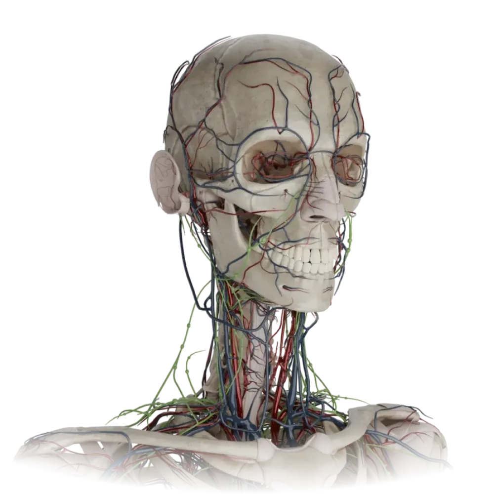
Evaluate the knowledge of the anatomy of the vessels of the head and neck. The test examines the topography, branching, anastomoses, and blood supply zones of the carotid arteries.
1. At what level does the bifurcation of the common carotid artery into the external and internal carotid arteries usually occur?
-
Upper border of the thyroid cartilage (level C4)
The bifurcation of the common carotid artery is normally located at the level of the upper border of the thyroid cartilage, corresponding to the fourth cervical vertebra (C4).
-
Lower border of the cricoid cartilage (level C6)
The bifurcation of the common carotid artery is normally located at the level of the upper border of the thyroid cartilage, corresponding to the fourth cervical vertebra (C4).
-
Level of the hyoid bone (level C3)
The bifurcation of the common carotid artery is normally located at the level of the upper border of the thyroid cartilage, corresponding to the fourth cervical vertebra (C4).
-
Angle of the mandible level
The bifurcation of the common carotid artery is normally located at the level of the upper border of the thyroid cartilage, corresponding to the fourth cervical vertebra (C4).
-
I find it difficult to answer
The bifurcation of the common carotid artery is normally located at the level of the upper border of the thyroid cartilage, corresponding to the fourth cervical vertebra (C4).
2. Which of the following arteries is NOT a branch of the external carotid artery?
-
Facial artery (a. facialis)
The ophthalmic artery is the first major branch of the internal carotid artery, originating after it exits the cavernous sinus.
-
Superior thyroid artery (a. thyroidea superior)
The ophthalmic artery is the first major branch of the internal carotid artery, originating after it exits the cavernous sinus.
-
Lingual artery (a. lingualis)
The ophthalmic artery is the first major branch of the internal carotid artery, originating after it exits the cavernous sinus.
-
Ophthalmic artery (a. ophthalmica)
The ophthalmic artery is the first major branch of the internal carotid artery, originating after it exits the cavernous sinus.
-
I find it difficult to answer
The ophthalmic artery is the first major branch of the internal carotid artery, originating after it exits the cavernous sinus.
3. Through which anatomical structure does the internal carotid artery penetrate into the cranial cavity?
-
Jugular foramen (foramen jugulare)
The internal carotid artery enters the temporal bone pyramid through the external opening of the carotid canal and exits into the cranial cavity through the internal opening.
-
Carotid canal (canalis caroticus)
The internal carotid artery enters the temporal bone pyramid through the external opening of the carotid canal and exits into the cranial cavity through the internal opening.
-
Foramen ovale
The internal carotid artery enters the temporal bone pyramid through the external opening of the carotid canal and exits into the cranial cavity through the internal opening.
-
Foramen spinosum
The internal carotid artery enters the temporal bone pyramid through the external opening of the carotid canal and exits into the cranial cavity through the internal opening.
-
I find it difficult to answer
The internal carotid artery enters the temporal bone pyramid through the external opening of the carotid canal and exits into the cranial cavity through the internal opening.
4. Which artery is a terminal branch of the external carotid artery?
-
Occipital artery
The external carotid artery divides into its terminal branches — the superficial temporal and maxillary arteries — within the parotid gland.
-
Ascending pharyngeal artery
The external carotid artery divides into its terminal branches — the superficial temporal and maxillary arteries — within the parotid gland.
-
Superficial temporal artery
The external carotid artery divides into its terminal branches — the superficial temporal and maxillary arteries — within the parotid gland.
-
Posterior auricular artery
The external carotid artery divides into its terminal branches — the superficial temporal and maxillary arteries — within the parotid gland.
-
I find it difficult to answer
The external carotid artery divides into its terminal branches — the superficial temporal and maxillary arteries — within the parotid gland.
5. Under which muscle does the lingual artery (a. lingualis) pass in the neck region?
-
Mylohyoid muscle (m. mylohyoideus)
The lingual artery is located in the submandibular triangle and passes medially (deeper) to the hyoglossus muscle.
-
Digastric muscle (m. digastricus)
The lingual artery is located in the submandibular triangle and passes medially (deeper) to the hyoglossus muscle.
-
Hyoglossus muscle (m. hyoglossus)
The lingual artery is located in the submandibular triangle and passes medially (deeper) to the hyoglossus muscle.
-
Sternocleidomastoid muscle (m. sternocleidomastoideus)
The lingual artery is located in the submandibular triangle and passes medially (deeper) to the hyoglossus muscle.
-
I find it difficult to answer
The lingual artery is located in the submandibular triangle and passes medially (deeper) to the hyoglossus muscle.
6. From which artery does the middle meningeal artery (a. meningea media) branch?
-
Maxillary artery (a. maxillaris)
The middle meningeal artery branches from the first (mandibular) part of the maxillary artery and enters the cranial cavity through the foramen spinosum.
-
Facial artery (a. facialis)
The middle meningeal artery branches from the first (mandibular) part of the maxillary artery and enters the cranial cavity through the foramen spinosum.
-
Superficial temporal artery (a. temporalis superficialis)
The middle meningeal artery branches from the first (mandibular) part of the maxillary artery and enters the cranial cavity through the foramen spinosum.
-
Occipital artery (a. occipitalis)
The middle meningeal artery branches from the first (mandibular) part of the maxillary artery and enters the cranial cavity through the foramen spinosum.
-
I find it difficult to answer
The middle meningeal artery branches from the first (mandibular) part of the maxillary artery and enters the cranial cavity through the foramen spinosum.
7. Where does the facial artery (a. facialis) bend over the lower border of the mandible?
-
Behind the ramus of the mandible
The facial artery appears on the face, bending over the edge of the mandible just in front of the attachment of the masseter muscle, where its pulsation can be palpated.
-
In front of the masseter muscle (m. masseter)
The facial artery appears on the face, bending over the edge of the mandible just in front of the attachment of the masseter muscle, where its pulsation can be palpated.
-
Medially to the medial pterygoid muscle
The facial artery appears on the face, bending over the edge of the mandible just in front of the attachment of the masseter muscle, where its pulsation can be palpated.
-
In the region of the mental symphysis
The facial artery appears on the face, bending over the edge of the mandible just in front of the attachment of the masseter muscle, where its pulsation can be palpated.
-
I find it difficult to answer
The facial artery appears on the face, bending over the edge of the mandible just in front of the attachment of the masseter muscle, where its pulsation can be palpated.
8. Which artery is a branch of the internal carotid artery in the cranial cavity?
-
Middle cerebral artery
The anterior choroidal artery branches from the cerebral part of the internal carotid artery before its bifurcation. The middle cerebral is a terminal branch, but the anterior choroidal is a direct lateral branch.
-
Posterior cerebral artery
The anterior choroidal artery branches from the cerebral part of the internal carotid artery before its bifurcation. The middle cerebral is a terminal branch, but the anterior choroidal is a direct lateral branch.
-
Superior cerebellar artery
The anterior choroidal artery branches from the cerebral part of the internal carotid artery before its bifurcation. The middle cerebral is a terminal branch, but the anterior choroidal is a direct lateral branch.
-
Anterior choroidal artery (a. choroidea anterior)
The anterior choroidal artery branches from the cerebral part of the internal carotid artery before its bifurcation. The middle cerebral is a terminal branch, but the anterior choroidal is a direct lateral branch.
-
I find it difficult to answer
The anterior choroidal artery branches from the cerebral part of the internal carotid artery before its bifurcation. The middle cerebral is a terminal branch, but the anterior choroidal is a direct lateral branch.
9. Which artery passes into the cranial cavity through the foramen spinosum (foramen spinosum)?
-
Middle meningeal artery
The middle meningeal artery, a branch of the maxillary artery, enters the middle cranial fossa through the foramen spinosum of the sphenoid bone.
-
Accessory meningeal artery
The middle meningeal artery, a branch of the maxillary artery, enters the middle cranial fossa through the foramen spinosum of the sphenoid bone.
-
Anterior meningeal artery
The middle meningeal artery, a branch of the maxillary artery, enters the middle cranial fossa through the foramen spinosum of the sphenoid bone.
-
Posterior meningeal artery
The middle meningeal artery, a branch of the maxillary artery, enters the middle cranial fossa through the foramen spinosum of the sphenoid bone.
-
I find it difficult to answer
The middle meningeal artery, a branch of the maxillary artery, enters the middle cranial fossa through the foramen spinosum of the sphenoid bone.
10. In which part of the internal carotid artery is its siphon located?
-
Cervical part (pars cervicalis)
The internal carotid artery forms an S-shaped bend (siphon) in its cavernous part, passing within the cavernous venous sinus.
-
Cavernous part (pars cavernosa)
The internal carotid artery forms an S-shaped bend (siphon) in its cavernous part, passing within the cavernous venous sinus.
-
Petrous part (pars petrosa)
The internal carotid artery forms an S-shaped bend (siphon) in its cavernous part, passing within the cavernous venous sinus.
-
Cerebral part (pars cerebralis)
The internal carotid artery forms an S-shaped bend (siphon) in its cavernous part, passing within the cavernous venous sinus.
-
I find it difficult to answer
The internal carotid artery forms an S-shaped bend (siphon) in its cavernous part, passing within the cavernous venous sinus.
11. The inferior alveolar artery (a. alveolaris inferior) is a branch of:
-
Facial artery
The inferior alveolar artery branches from the first part of the maxillary artery and enters the mandibular canal.
-
Lingual artery
The inferior alveolar artery branches from the first part of the maxillary artery and enters the mandibular canal.
-
Maxillary artery
The inferior alveolar artery branches from the first part of the maxillary artery and enters the mandibular canal.
-
Superficial temporal artery
The inferior alveolar artery branches from the first part of the maxillary artery and enters the mandibular canal.
-
I find it difficult to answer
The inferior alveolar artery branches from the first part of the maxillary artery and enters the mandibular canal.
12. Which artery predominantly supplies the tympanic cavity and auricle?
-
Facial artery
The posterior auricular artery branches from the posterior surface of the external carotid artery and gives branches to the auricle and tympanic cavity (styloid-mastoid artery).
-
Lingual artery
The posterior auricular artery branches from the posterior surface of the external carotid artery and gives branches to the auricle and tympanic cavity (styloid-mastoid artery).
-
Ascending pharyngeal artery
The posterior auricular artery branches from the posterior surface of the external carotid artery and gives branches to the auricle and tympanic cavity (styloid-mastoid artery).
-
Posterior auricular artery (a. auricularis posterior)
The posterior auricular artery branches from the posterior surface of the external carotid artery and gives branches to the auricle and tympanic cavity (styloid-mastoid artery).
-
I find it difficult to answer
The posterior auricular artery branches from the posterior surface of the external carotid artery and gives branches to the auricle and tympanic cavity (styloid-mastoid artery).
13. From which surface of the external carotid artery does the superior thyroid artery branch?
-
From the anterior surface
The superior thyroid, lingual, and facial arteries form the group of anterior branches of the external carotid artery.
-
From the posterior surface
The superior thyroid, lingual, and facial arteries form the group of anterior branches of the external carotid artery.
-
From the medial surface
The superior thyroid, lingual, and facial arteries form the group of anterior branches of the external carotid artery.
-
From the lateral surface
The superior thyroid, lingual, and facial arteries form the group of anterior branches of the external carotid artery.
-
I find it difficult to answer
The superior thyroid, lingual, and facial arteries form the group of anterior branches of the external carotid artery.
14. In which groove does the occipital artery (a. occipitalis) lie?
-
Groove for sigmoid sinus
The occipital artery lies in its corresponding groove (sulcus arteriae occipitalis), located medially to the mastoid notch of the temporal bone.
-
Groove for the occipital artery on the mastoid process
The occipital artery lies in its corresponding groove (sulcus arteriae occipitalis), located medially to the mastoid notch of the temporal bone.
-
Groove for transverse sinus
The occipital artery lies in its corresponding groove (sulcus arteriae occipitalis), located medially to the mastoid notch of the temporal bone.
-
Carotid groove of the sphenoid bone
The occipital artery lies in its corresponding groove (sulcus arteriae occipitalis), located medially to the mastoid notch of the temporal bone.
-
I find it difficult to answer
The occipital artery lies in its corresponding groove (sulcus arteriae occipitalis), located medially to the mastoid notch of the temporal bone.
15. The ascending pharyngeal artery (a. pharyngea ascendens) arises from:
-
The anterior surface of the external carotid artery
The ascending pharyngeal artery is the only medial branch of the external carotid artery, originating deeply from its beginning.
-
External carotid artery in the region of its bifurcation
The ascending pharyngeal artery is the only medial branch of the external carotid artery, originating deeply from its beginning.
-
Medial semicircle of the external carotid artery
The ascending pharyngeal artery is the only medial branch of the external carotid artery, originating deeply from its beginning.
-
Internal carotid artery
The ascending pharyngeal artery is the only medial branch of the external carotid artery, originating deeply from its beginning.
-
I find it difficult to answer
The ascending pharyngeal artery is the only medial branch of the external carotid artery, originating deeply from its beginning.
16. With which artery does the dorsal nasal artery (a branch of the ophthalmic artery) anastomose?
-
With the supratrochlear artery
The angular artery (the terminal branch of the facial artery) anastomoses with the dorsal nasal artery at the medial angle of the eye, forming a connection between the systems of the external and internal carotid arteries.
-
With the infraorbital artery
The angular artery (the terminal branch of the facial artery) anastomoses with the dorsal nasal artery at the medial angle of the eye, forming a connection between the systems of the external and internal carotid arteries.
-
With the transverse facial artery
The angular artery (the terminal branch of the facial artery) anastomoses with the dorsal nasal artery at the medial angle of the eye, forming a connection between the systems of the external and internal carotid arteries.
-
With the angular artery (a. angularis)
The angular artery (the terminal branch of the facial artery) anastomoses with the dorsal nasal artery at the medial angle of the eye, forming a connection between the systems of the external and internal carotid arteries.
-
I find it difficult to answer
The angular artery (the terminal branch of the facial artery) anastomoses with the dorsal nasal artery at the medial angle of the eye, forming a connection between the systems of the external and internal carotid arteries.
17. The sphenopalatine artery (a. sphenopalatina) is a terminal branch of:
-
Maxillary artery
The sphenopalatine artery branches out from the third (pterygopalatine) part of the maxillary artery and enters the nasal cavity.
-
Facial artery
The sphenopalatine artery branches out from the third (pterygopalatine) part of the maxillary artery and enters the nasal cavity.
-
Ophthalmic artery
The sphenopalatine artery branches out from the third (pterygopalatine) part of the maxillary artery and enters the nasal cavity.
-
Internal carotid artery
The sphenopalatine artery branches out from the third (pterygopalatine) part of the maxillary artery and enters the nasal cavity.
-
I find it difficult to answer
The sphenopalatine artery branches out from the third (pterygopalatine) part of the maxillary artery and enters the nasal cavity.
18. The posterior communicating artery (a. communicans posterior) connects the internal carotid artery to:
-
Anterior cerebral artery
The posterior communicating artery originates from the internal carotid artery and flows into the posterior cerebral artery, contributing to the formation of the circle of Willis.
-
Posterior cerebral artery
The posterior communicating artery originates from the internal carotid artery and flows into the posterior cerebral artery, contributing to the formation of the circle of Willis.
-
Middle cerebral artery
The posterior communicating artery originates from the internal carotid artery and flows into the posterior cerebral artery, contributing to the formation of the circle of Willis.
-
Basilar artery
The posterior communicating artery originates from the internal carotid artery and flows into the posterior cerebral artery, contributing to the formation of the circle of Willis.
-
I find it difficult to answer
The posterior communicating artery originates from the internal carotid artery and flows into the posterior cerebral artery, contributing to the formation of the circle of Willis.
19. In which topographic structure of the neck is the lingual artery located (Pirogov's triangle)?
-
Carotid triangle
The lingual triangle (Pirogov's triangle) is part of the submandibular triangle of the neck, where the lingual artery is accessible for ligation.
-
Omotracheal triangle
The lingual triangle (Pirogov's triangle) is part of the submandibular triangle of the neck, where the lingual artery is accessible for ligation.
-
Submandibular triangle
The lingual triangle (Pirogov's triangle) is part of the submandibular triangle of the neck, where the lingual artery is accessible for ligation.
-
Occipital triangle
The lingual triangle (Pirogov's triangle) is part of the submandibular triangle of the neck, where the lingual artery is accessible for ligation.
-
I find it difficult to answer
The lingual triangle (Pirogov's triangle) is part of the submandibular triangle of the neck, where the lingual artery is accessible for ligation.
20. Which artery supplies the dura mater of the anterior cranial fossa (anterior meningeal artery)?
-
Middle meningeal artery
The anterior meningeal branch originates from the anterior ethmoidal artery (a branch of the ophthalmic artery from the internal carotid system) and supplies the dura mater of the anterior cranial fossa.
-
Ascending pharyngeal artery
The anterior meningeal branch originates from the anterior ethmoidal artery (a branch of the ophthalmic artery from the internal carotid system) and supplies the dura mater of the anterior cranial fossa.
-
Occipital artery
The anterior meningeal branch originates from the anterior ethmoidal artery (a branch of the ophthalmic artery from the internal carotid system) and supplies the dura mater of the anterior cranial fossa.
-
Anterior ethmoidal artery (a. ethmoidalis anterior)
The anterior meningeal branch originates from the anterior ethmoidal artery (a branch of the ophthalmic artery from the internal carotid system) and supplies the dura mater of the anterior cranial fossa.
-
I find it difficult to answer
The anterior meningeal branch originates from the anterior ethmoidal artery (a branch of the ophthalmic artery from the internal carotid system) and supplies the dura mater of the anterior cranial fossa.
Retake this quiz?
Your current progress will be reset.
Vessels of the head and neck
Internal carotid artery
0/20