


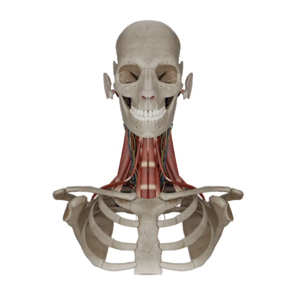
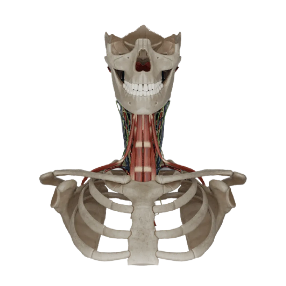
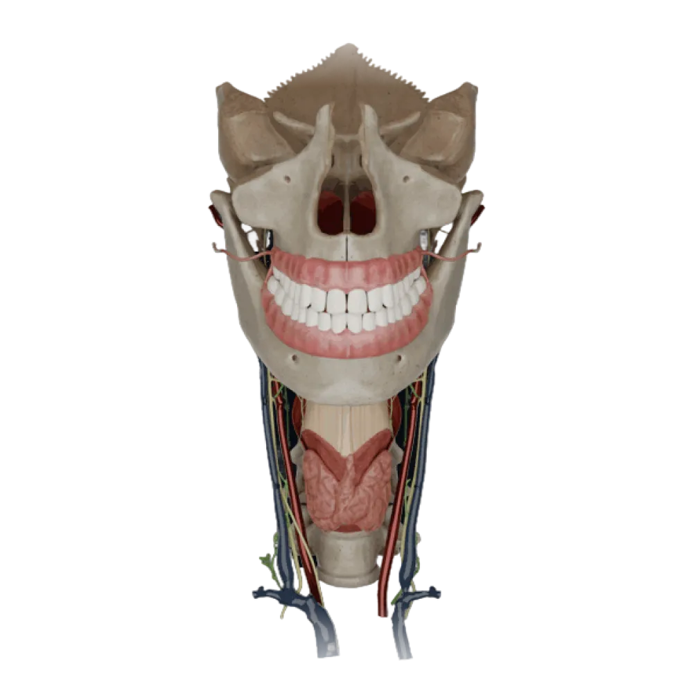
Pharyngeal Anatomy Test
Check your knowledge of pharyngeal anatomy. The test covers skeletal landmarks, topography, muscles, blood supply, lymphatic drainage, and innervation of the complex.
1/20
bold
text
1. Specify the skeletal landmarks of the pharynx in adults.
-
From the cranial base to the VI-VII cervical vertebrae
In adults, the pharynx begins at the cranial base and transitions into the esophagus at the level of the lower edge of the VI cervical vertebra.
-
From the cranial base to the IV thoracic vertebra
In adults, the pharynx begins at the cranial base and transitions into the esophagus at the level of the lower edge of the VI cervical vertebra.
-
From the hard palate to the V cervical vertebra
In adults, the pharynx begins at the cranial base and transitions into the esophagus at the level of the lower edge of the VI cervical vertebra.
-
From the hyoid bone to the jugular notch of the sternum
In adults, the pharynx begins at the cranial base and transitions into the esophagus at the level of the lower edge of the VI cervical vertebra.
-
I find it difficult to answer
In adults, the pharynx begins at the cranial base and transitions into the esophagus at the level of the lower edge of the VI cervical vertebra.
2. Which nerves participate in the formation of the pharyngeal plexus (plexus pharyngeus)?
-
Trigeminal, facial, and vagus nerves
The pharyngeal plexus is formed by branches of the glossopharyngeal (IX), vagus (X) nerves and the sympathetic trunk.
-
Glossopharyngeal, vagus nerves, and sympathetic fibers
The pharyngeal plexus is formed by branches of the glossopharyngeal (IX), vagus (X) nerves and the sympathetic trunk.
-
Hypoglossal, glossopharyngeal, and accessory nerves
The pharyngeal plexus is formed by branches of the glossopharyngeal (IX), vagus (X) nerves and the sympathetic trunk.
-
Facial, glossopharyngeal nerves, and parasympathetic fibers
The pharyngeal plexus is formed by branches of the glossopharyngeal (IX), vagus (X) nerves and the sympathetic trunk.
-
I find it difficult to answer
The pharyngeal plexus is formed by branches of the glossopharyngeal (IX), vagus (X) nerves and the sympathetic trunk.
3. Which artery is the primary source of blood supply to the pharynx?
-
Descending palatine artery
The ascending pharyngeal artery (a. pharyngea ascendens), a branch of the external carotid artery, is the primary source of blood supply to the pharynx.
-
Lingual artery
The ascending pharyngeal artery (a. pharyngea ascendens), a branch of the external carotid artery, is the primary source of blood supply to the pharynx.
-
Ascending pharyngeal artery
The ascending pharyngeal artery (a. pharyngea ascendens), a branch of the external carotid artery, is the primary source of blood supply to the pharynx.
-
Superior laryngeal artery
The ascending pharyngeal artery (a. pharyngea ascendens), a branch of the external carotid artery, is the primary source of blood supply to the pharynx.
-
I find it difficult to answer
The ascending pharyngeal artery (a. pharyngea ascendens), a branch of the external carotid artery, is the primary source of blood supply to the pharynx.
4. Which nerve provides motor innervation to the stylopharyngeus muscle?
-
Vagus nerve
The stylopharyngeus muscle is the only pharyngeal muscle innervated by the glossopharyngeal nerve (CN IX); the others are innervated by the vagus nerve.
-
Accessory nerve
The stylopharyngeus muscle is the only pharyngeal muscle innervated by the glossopharyngeal nerve (CN IX); the others are innervated by the vagus nerve.
-
Trigeminal nerve.
The stylopharyngeus muscle is the only pharyngeal muscle innervated by the glossopharyngeal nerve (CN IX); the others are innervated by the vagus nerve.
-
Glossopharyngeal nerve
The stylopharyngeus muscle is the only pharyngeal muscle innervated by the glossopharyngeal nerve (CN IX); the others are innervated by the vagus nerve.
-
I find it difficult to answer
The stylopharyngeus muscle is the only pharyngeal muscle innervated by the glossopharyngeal nerve (CN IX); the others are innervated by the vagus nerve.
5. Where is the piriform recess (recessus piriformis) located?
-
In the nasal portion of the pharynx, posterior to the tubal fold
The piriform recess is a depression in the laryngeal part of the pharynx, located laterally to the entrance of the larynx.
-
In the oral portion of the pharynx, between the palatine arches
The piriform recess is a depression in the laryngeal part of the pharynx, located laterally to the entrance of the larynx.
-
In the laryngeal part of the pharynx, on the sides of the laryngeal inlet
The piriform recess is a depression in the laryngeal part of the pharynx, located laterally to the entrance of the larynx.
-
At the transition point from the pharynx to the esophagus
The piriform recess is a depression in the laryngeal part of the pharynx, located laterally to the entrance of the larynx.
-
I find it difficult to answer
The piriform recess is a depression in the laryngeal part of the pharynx, located laterally to the entrance of the larynx.
6. To which structure is the pharyngobasilar fascia attached at the cranial base?
-
To the pharyngeal tubercle of the occipital bone
The pharyngobasilar fascia is attached to the pharyngeal tubercle of the basilar part of the occipital bone, the pyramid of the temporal bone, and the medial plate of the pterygoid process.
-
To the foramen magnum
The pharyngobasilar fascia is attached to the pharyngeal tubercle of the basilar part of the occipital bone, the pyramid of the temporal bone, and the medial plate of the pterygoid process.
-
To the styloid process of the temporal bone
The pharyngobasilar fascia is attached to the pharyngeal tubercle of the basilar part of the occipital bone, the pyramid of the temporal bone, and the medial plate of the pterygoid process.
-
To the mastoid process
The pharyngobasilar fascia is attached to the pharyngeal tubercle of the basilar part of the occipital bone, the pyramid of the temporal bone, and the medial plate of the pterygoid process.
-
I find it difficult to answer
The pharyngobasilar fascia is attached to the pharyngeal tubercle of the basilar part of the occipital bone, the pyramid of the temporal bone, and the medial plate of the pterygoid process.
7. By which anatomical formations does the nasal part of the pharynx communicate with the nasal cavity?
-
Isthmus
The nasal part of the pharynx communicates with the nasal cavity anteriorly through paired openings called choanae.
-
Choanae
The nasal part of the pharynx communicates with the nasal cavity anteriorly through paired openings called choanae.
-
Pharyngeal openings of the auditory tubes
The nasal part of the pharynx communicates with the nasal cavity anteriorly through paired openings called choanae.
-
Piriform apertures
The nasal part of the pharynx communicates with the nasal cavity anteriorly through paired openings called choanae.
-
I find it difficult to answer
The nasal part of the pharynx communicates with the nasal cavity anteriorly through paired openings called choanae.
8. On which wall of the pharynx is the pharyngeal opening of the auditory (Eustachian) tube located?
-
On the anterior wall of the oral part
The pharyngeal opening of the auditory tube (ostium pharyngeum tubae auditivae) is located on the lateral wall of the nasopharynx at the level of the inferior turbinate.
-
On the posterior wall of the laryngeal part
The pharyngeal opening of the auditory tube (ostium pharyngeum tubae auditivae) is located on the lateral wall of the nasopharynx at the level of the inferior turbinate.
-
On the superior wall of the nasal part
The pharyngeal opening of the auditory tube (ostium pharyngeum tubae auditivae) is located on the lateral wall of the nasopharynx at the level of the inferior turbinate.
-
On the lateral wall of the nasal part
The pharyngeal opening of the auditory tube (ostium pharyngeum tubae auditivae) is located on the lateral wall of the nasopharynx at the level of the inferior turbinate.
-
I find it difficult to answer
The pharyngeal opening of the auditory tube (ostium pharyngeum tubae auditivae) is located on the lateral wall of the nasopharynx at the level of the inferior turbinate.
9. Where do the tendinous bundles of the pharyngeal constrictors of opposite sides join?
-
On the hyoid bone
The fibers of all three pharyngeal constrictors converge on the posterior surface and intertwine into the tendinous strip — the pharyngeal raphe (raphe pharyngis).
-
On the thyroid cartilage
The fibers of all three pharyngeal constrictors converge on the posterior surface and intertwine into the tendinous strip — the pharyngeal raphe (raphe pharyngis).
-
Along the median line of the posterior wall of the pharynx, forming the pharyngeal raphe
The fibers of all three pharyngeal constrictors converge on the posterior surface and intertwine into the tendinous strip — the pharyngeal raphe (raphe pharyngis).
-
On the pharyngobasilar fascia
The fibers of all three pharyngeal constrictors converge on the posterior surface and intertwine into the tendinous strip — the pharyngeal raphe (raphe pharyngis).
-
I find it difficult to answer
The fibers of all three pharyngeal constrictors converge on the posterior surface and intertwine into the tendinous strip — the pharyngeal raphe (raphe pharyngis).
10. Into which lymph nodes does lymph predominantly drain from the upper part of the pharynx?
-
Submental lymph nodes
Lymph from the pharynx drains into the retropharyngeal nodes (nodi lymphatici retropharyngeales) and the deep lateral cervical nodes along the internal jugular vein.
-
Axillary lymph nodes
Lymph from the pharynx drains into the retropharyngeal nodes (nodi lymphatici retropharyngeales) and the deep lateral cervical nodes along the internal jugular vein.
-
Superficial cervical lymph nodes
Lymph from the pharynx drains into the retropharyngeal nodes (nodi lymphatici retropharyngeales) and the deep lateral cervical nodes along the internal jugular vein.
-
Retropharyngeal and deep cervical lymph nodes
Lymph from the pharynx drains into the retropharyngeal nodes (nodi lymphatici retropharyngeales) and the deep lateral cervical nodes along the internal jugular vein.
-
I find it difficult to answer
Lymph from the pharynx drains into the retropharyngeal nodes (nodi lymphatici retropharyngeales) and the deep lateral cervical nodes along the internal jugular vein.
11. Which muscles are included in the group of pharyngeal elevators (longitudinal muscles)?
-
Superior, middle, and inferior constrictors
The longitudinal muscles that elevate and expand the pharynx include m. stylopharyngeus, m. palatopharyngeus and m. salpingopharyngeus.
-
Stylopharyngeus, palatopharyngeus, and salpingopharyngeus muscles
The longitudinal muscles that elevate and expand the pharynx include m. stylopharyngeus, m. palatopharyngeus and m. salpingopharyngeus.
-
Mylohyoid and digastric muscles
The longitudinal muscles that elevate and expand the pharynx include m. stylopharyngeus, m. palatopharyngeus and m. salpingopharyngeus.
-
Styloglossus and hyoglossus muscles
The longitudinal muscles that elevate and expand the pharynx include m. stylopharyngeus, m. palatopharyngeus and m. salpingopharyngeus.
-
I find it difficult to answer
The longitudinal muscles that elevate and expand the pharynx include m. stylopharyngeus, m. palatopharyngeus and m. salpingopharyngeus.
12. What tonsils, along with the palatine and lingual, form the pharyngeal lymphoid ring (Waldeyer's ring)?
-
Pharyngeal and tubal tonsils
Pirogov-Waldeyer's lymphoepithelial ring includes one lingual, one pharyngeal, two palatine, and two tubal tonsils.
-
Laryngeal and tracheal tonsils
Pirogov-Waldeyer's lymphoepithelial ring includes one lingual, one pharyngeal, two palatine, and two tubal tonsils.
-
Pharyngeal and laryngeal tonsils
Pirogov-Waldeyer's lymphoepithelial ring includes one lingual, one pharyngeal, two palatine, and two tubal tonsils.
-
Tubal and buccal tonsils
Pirogov-Waldeyer's lymphoepithelial ring includes one lingual, one pharyngeal, two palatine, and two tubal tonsils.
-
I find it difficult to answer
Pirogov-Waldeyer's lymphoepithelial ring includes one lingual, one pharyngeal, two palatine, and two tubal tonsils.
13. Where does the main venous drainage from the pharyngeal venous plexus flow?
-
Into the external jugular vein.
The pharyngeal venous plexus collects blood from the pharyngeal walls and drains through the pharyngeal veins into the internal jugular vein.
-
Into the facial vein.
The pharyngeal venous plexus collects blood from the pharyngeal walls and drains through the pharyngeal veins into the internal jugular vein.
-
Into the vertebral vein
The pharyngeal venous plexus collects blood from the pharyngeal walls and drains through the pharyngeal veins into the internal jugular vein.
-
Into the internal jugular vein.
The pharyngeal venous plexus collects blood from the pharyngeal walls and drains through the pharyngeal veins into the internal jugular vein.
-
I find it difficult to answer
The pharyngeal venous plexus collects blood from the pharyngeal walls and drains through the pharyngeal veins into the internal jugular vein.
14. Which nerve branches provide sensory innervation to the mucous membrane of the nasal part of the pharynx?
-
Maxillary nerve (V2)
The mucous membrane of the superior part of the pharynx (nasopharynx) is innervated by pharyngeal branches of the pterygopalatine ganglion, related to the maxillary nerve.
-
Glossopharyngeal nerve (IX)
The mucous membrane of the superior part of the pharynx (nasopharynx) is innervated by pharyngeal branches of the pterygopalatine ganglion, related to the maxillary nerve.
-
Vagus nerve (X)
The mucous membrane of the superior part of the pharynx (nasopharynx) is innervated by pharyngeal branches of the pterygopalatine ganglion, related to the maxillary nerve.
-
Facial nerve (VII)
The mucous membrane of the superior part of the pharynx (nasopharynx) is innervated by pharyngeal branches of the pterygopalatine ganglion, related to the maxillary nerve.
-
I find it difficult to answer
The mucous membrane of the superior part of the pharynx (nasopharynx) is innervated by pharyngeal branches of the pterygopalatine ganglion, related to the maxillary nerve.
15. What anatomical structure is located anteriorly to the oral part of the pharynx?
-
Larynx
The oral part of the pharynx extends from the level of the soft palate to the entrance of the larynx, and anteriorly it communicates with the oral cavity through the isthmus.
-
Nasal cavity
The oral part of the pharynx extends from the level of the soft palate to the entrance of the larynx, and anteriorly it communicates with the oral cavity through the isthmus.
-
Isthmus and root of the tongue
The oral part of the pharynx extends from the level of the soft palate to the entrance of the larynx, and anteriorly it communicates with the oral cavity through the isthmus.
-
Esophagus
The oral part of the pharynx extends from the level of the soft palate to the entrance of the larynx, and anteriorly it communicates with the oral cavity through the isthmus.
-
I find it difficult to answer
The oral part of the pharynx extends from the level of the soft palate to the entrance of the larynx, and anteriorly it communicates with the oral cavity through the isthmus.
16. From which structures does the middle pharyngeal constrictor originate?
-
From the medial plate of the pterygoid process and the mylohyoid line
The middle pharyngeal constrictor (m. constrictor pharyngis medius) originates from the greater and lesser horns of the hyoid bone and the stylohyoid ligament.
-
From the greater and lesser horns of the hyoid bone
The middle pharyngeal constrictor (m. constrictor pharyngis medius) originates from the greater and lesser horns of the hyoid bone and the stylohyoid ligament.
-
From the thyroid and cricoid cartilages of the larynx
The middle pharyngeal constrictor (m. constrictor pharyngis medius) originates from the greater and lesser horns of the hyoid bone and the stylohyoid ligament.
-
From the styloid process of the temporal bone
The middle pharyngeal constrictor (m. constrictor pharyngis medius) originates from the greater and lesser horns of the hyoid bone and the stylohyoid ligament.
-
I find it difficult to answer
The middle pharyngeal constrictor (m. constrictor pharyngis medius) originates from the greater and lesser horns of the hyoid bone and the stylohyoid ligament.
17. What limits the retropharyngeal space (spatium retropharyngeum) posteriorly?
-
Buccopharyngeal fascia
The retropharyngeal space is located between the posterior wall of the pharynx anteriorly and the prevertebral layer of cervical fascia posteriorly.
-
Pharyngobasilar fascia
The retropharyngeal space is located between the posterior wall of the pharynx anteriorly and the prevertebral layer of cervical fascia posteriorly.
-
Mucosa of the pharynx
The retropharyngeal space is located between the posterior wall of the pharynx anteriorly and the prevertebral layer of cervical fascia posteriorly.
-
Prevertebral layer of cervical fascia
The retropharyngeal space is located between the posterior wall of the pharynx anteriorly and the prevertebral layer of cervical fascia posteriorly.
-
I find it difficult to answer
The retropharyngeal space is located between the posterior wall of the pharynx anteriorly and the prevertebral layer of cervical fascia posteriorly.
18. What two parts can be distinguished in the inferior pharyngeal constrictor?
-
Pterygo-pharyngeal and bucco-pharyngeal
The inferior pharyngeal constrictor has two parts, named after their origins: thyro-pharyngeal (pars thyropharyngea) and crico-pharyngeal (pars cricopharyngea).
-
Chondro-pharyngeal and cornu-pharyngeal
The inferior pharyngeal constrictor has two parts, named after their origins: thyro-pharyngeal (pars thyropharyngea) and crico-pharyngeal (pars cricopharyngea).
-
Thyro-pharyngeal and crico-pharyngeal
The inferior pharyngeal constrictor has two parts, named after their origins: thyro-pharyngeal (pars thyropharyngea) and crico-pharyngeal (pars cricopharyngea).
-
Maxillo-pharyngeal and glossopharyngeal
The inferior pharyngeal constrictor has two parts, named after their origins: thyro-pharyngeal (pars thyropharyngea) and crico-pharyngeal (pars cricopharyngea).
-
I find it difficult to answer
The inferior pharyngeal constrictor has two parts, named after their origins: thyro-pharyngeal (pars thyropharyngea) and crico-pharyngeal (pars cricopharyngea).
19. What structure forms the base of the torus tubarius on the lateral wall of the pharynx?
-
Cartilaginous part of the auditory tube
The torus tubarius is formed by the medial cartilaginous edge of the auditory (Eustachian) tube protruding into the nasopharynx.
-
Medial pterygoid plate
The torus tubarius is formed by the medial cartilaginous edge of the auditory (Eustachian) tube protruding into the nasopharynx.
-
Lymphoid tissue of the pharyngeal tonsil
The torus tubarius is formed by the medial cartilaginous edge of the auditory (Eustachian) tube protruding into the nasopharynx.
-
Tube-pharyngeal muscle
The torus tubarius is formed by the medial cartilaginous edge of the auditory (Eustachian) tube protruding into the nasopharynx.
-
I find it difficult to answer
The torus tubarius is formed by the medial cartilaginous edge of the auditory (Eustachian) tube protruding into the nasopharynx.
20. In what order (from the inside out) are the layers of the pharyngeal wall positioned in its upper region?
-
Mucosa, muscular, pharyngo-basilar fascia, adventitia
The inside of the wall is lined by a mucous membrane, outside of which lies a fibrous base (pharyngo-basilar fascia), followed by a muscular layer covered by adventitia.
-
Mucosa, pharyngo-basilar fascia, muscular, adventitia
The inside of the wall is lined by a mucous membrane, outside of which lies a fibrous base (pharyngo-basilar fascia), followed by a muscular layer covered by adventitia.
-
Muscular, mucosa, adventitia, serous membrane
The inside of the wall is lined by a mucous membrane, outside of which lies a fibrous base (pharyngo-basilar fascia), followed by a muscular layer covered by adventitia.
-
Mucosa, submucosa, muscular, serous membrane
The inside of the wall is lined by a mucous membrane, outside of which lies a fibrous base (pharyngo-basilar fascia), followed by a muscular layer covered by adventitia.
-
I find it difficult to answer
The inside of the wall is lined by a mucous membrane, outside of which lies a fibrous base (pharyngo-basilar fascia), followed by a muscular layer covered by adventitia.
Retake this quiz?
Your current progress will be reset.