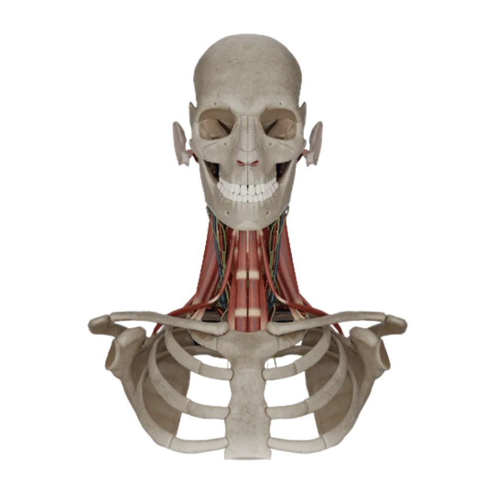
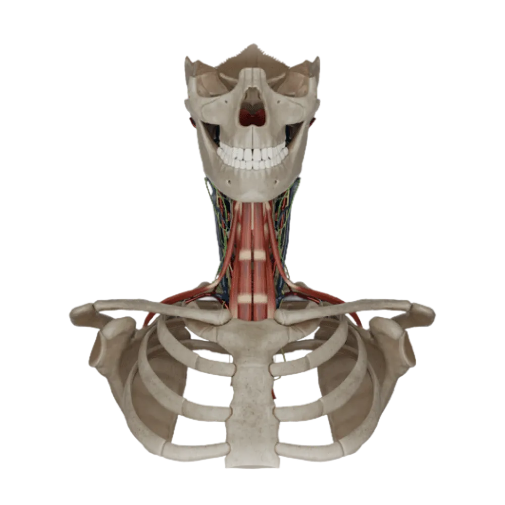
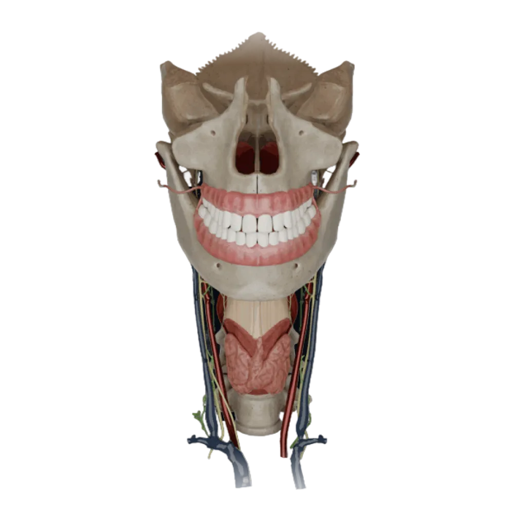
Check your knowledge of head and neck organ anatomy. The test assesses the topography, blood supply, and innervation of the pharynx, larynx, glands, eye, and ear.
1/20
0%
bold
text
1. Taste innervation of the anterior two-thirds of the tongue is provided by the branch of:
Glossopharyngeal nerve.
The chorda tympani, a branch of the facial nerve (VII), specifically provides taste innervation to the anterior 2/3 of the tongue.
Vagus nerve.
The chorda tympani, a branch of the facial nerve (VII), specifically provides taste innervation to the anterior 2/3 of the tongue.
Chorda tympani (from the facial nerve).
The chorda tympani, a branch of the facial nerve (VII), specifically provides taste innervation to the anterior 2/3 of the tongue.
Lingual nerve (from the trigeminal nerve).
The chorda tympani, a branch of the facial nerve (VII), specifically provides taste innervation to the anterior 2/3 of the tongue.
I find it difficult to answer
The chorda tympani, a branch of the facial nerve (VII), specifically provides taste innervation to the anterior 2/3 of the tongue.
2. Which muscle, when contracted bilaterally, protrudes the tongue forward?
Genioglossus muscle.
The genioglossus muscle (m. genioglossus) when bilaterally contracted pulls the tongue forward and down.
Hyoglossus muscle.
The genioglossus muscle (m. genioglossus) when bilaterally contracted pulls the tongue forward and down.
Styloglossus muscle.
The genioglossus muscle (m. genioglossus) when bilaterally contracted pulls the tongue forward and down.
Palatoglossus muscle.
The genioglossus muscle (m. genioglossus) when bilaterally contracted pulls the tongue forward and down.
I find it difficult to answer
The genioglossus muscle (m. genioglossus) when bilaterally contracted pulls the tongue forward and down.
3. The main arterial source of blood supply to the upper part of the pharynx is:
Descending palatine artery
The ascending pharyngeal artery (a. pharyngea ascendens), a branch of the external carotid artery, is the main source of blood supply to the upper parts of the pharynx.
Ascending pharyngeal artery
The ascending pharyngeal artery (a. pharyngea ascendens), a branch of the external carotid artery, is the main source of blood supply to the upper parts of the pharynx.
Superior thyroid artery
The ascending pharyngeal artery (a. pharyngea ascendens), a branch of the external carotid artery, is the main source of blood supply to the upper parts of the pharynx.
Facial artery
The ascending pharyngeal artery (a. pharyngea ascendens), a branch of the external carotid artery, is the main source of blood supply to the upper parts of the pharynx.
I find it difficult to answer
The ascending pharyngeal artery (a. pharyngea ascendens), a branch of the external carotid artery, is the main source of blood supply to the upper parts of the pharynx.
4. At the level of which cervical vertebra does the pharynx transition into the esophagus in adults?
C4
In adults, the lower boundary of the pharynx and its transition into the esophagus projects at the level of the lower edge of the cricoid cartilage, corresponding to the sixth cervical vertebra (C6).
C5
In adults, the lower boundary of the pharynx and its transition into the esophagus projects at the level of the lower edge of the cricoid cartilage, corresponding to the sixth cervical vertebra (C6).
C7
In adults, the lower boundary of the pharynx and its transition into the esophagus projects at the level of the lower edge of the cricoid cartilage, corresponding to the sixth cervical vertebra (C6).
C6
In adults, the lower boundary of the pharynx and its transition into the esophagus projects at the level of the lower edge of the cricoid cartilage, corresponding to the sixth cervical vertebra (C6).
I find it difficult to answer
In adults, the lower boundary of the pharynx and its transition into the esophagus projects at the level of the lower edge of the cricoid cartilage, corresponding to the sixth cervical vertebra (C6).
5. Where does the aperture of the sphenoid sinus (sinus sphenoidalis) open into?
Into the superior nasal meatus.
The sphenoid sinus opens its aperture into the sphenoethmoidal recess, located above and posterior to the superior turbinate.
Into the sphenoethmoidal recess.
The sphenoid sinus opens its aperture into the sphenoethmoidal recess, located above and posterior to the superior turbinate.
Into the middle nasal meatus.
The sphenoid sinus opens its aperture into the sphenoethmoidal recess, located above and posterior to the superior turbinate.
Into the inferior nasal meatus.
The sphenoid sinus opens its aperture into the sphenoethmoidal recess, located above and posterior to the superior turbinate.
I find it difficult to answer
The sphenoid sinus opens its aperture into the sphenoethmoidal recess, located above and posterior to the superior turbinate.
6. The primary source of the sphenopalatine artery (a. sphenopalatina), supplying the nasal cavity, is:
Maxillary artery
The sphenopalatine artery is the terminal branch of the maxillary artery (a. maxillaris) from the external carotid artery system.
Facial artery
The sphenopalatine artery is the terminal branch of the maxillary artery (a. maxillaris) from the external carotid artery system.
Ophthalmic artery
The sphenopalatine artery is the terminal branch of the maxillary artery (a. maxillaris) from the external carotid artery system.
Superficial temporal artery
The sphenopalatine artery is the terminal branch of the maxillary artery (a. maxillaris) from the external carotid artery system.
I find it difficult to answer
The sphenopalatine artery is the terminal branch of the maxillary artery (a. maxillaris) from the external carotid artery system.
7. Which laryngeal muscle is the sole dilator of the glottis (abducts the vocal folds)?
Lateral cricoarytenoid muscle.
The posterior cricoarytenoid muscle (m. cricoarytenoideus posterior) rotates the arytenoid cartilages during contraction, expanding the glottis.
Transverse arytenoid muscle
The posterior cricoarytenoid muscle (m. cricoarytenoideus posterior) rotates the arytenoid cartilages during contraction, expanding the glottis.
Posterior cricoarytenoid muscle.
The posterior cricoarytenoid muscle (m. cricoarytenoideus posterior) rotates the arytenoid cartilages during contraction, expanding the glottis.
Thyroarytenoid muscle.
The posterior cricoarytenoid muscle (m. cricoarytenoideus posterior) rotates the arytenoid cartilages during contraction, expanding the glottis.
I find it difficult to answer
The posterior cricoarytenoid muscle (m. cricoarytenoideus posterior) rotates the arytenoid cartilages during contraction, expanding the glottis.
8. The motor innervation of the cricothyroid muscle (m. cricothyroideus) is provided by:
By the internal branch of the superior laryngeal nerve.
Unlike the other laryngeal muscles, the cricothyroid muscle is innervated by the external branch of the superior laryngeal nerve (r. externus n. laryngei superioris).
Recurrent laryngeal nerve.
Unlike the other laryngeal muscles, the cricothyroid muscle is innervated by the external branch of the superior laryngeal nerve (r. externus n. laryngei superioris).
Glossopharyngeal nerve.
Unlike the other laryngeal muscles, the cricothyroid muscle is innervated by the external branch of the superior laryngeal nerve (r. externus n. laryngei superioris).
By the external branch of the superior laryngeal nerve.
Unlike the other laryngeal muscles, the cricothyroid muscle is innervated by the external branch of the superior laryngeal nerve (r. externus n. laryngei superioris).
I find it difficult to answer
Unlike the other laryngeal muscles, the cricothyroid muscle is innervated by the external branch of the superior laryngeal nerve (r. externus n. laryngei superioris).
9. Which nerve is topographically located in the tracheoesophageal groove, immediately medial to the lateral lobes of the thyroid gland?
Phrenic nerve
The recurrent laryngeal nerve (n. laryngeus recurrens) ascends to the larynx in the tracheoesophageal groove, closely adjacent to the posteromedial surface of the lobes of the thyroid gland.
Recurrent laryngeal nerve.
The recurrent laryngeal nerve (n. laryngeus recurrens) ascends to the larynx in the tracheoesophageal groove, closely adjacent to the posteromedial surface of the lobes of the thyroid gland.
Superior laryngeal nerve.
The recurrent laryngeal nerve (n. laryngeus recurrens) ascends to the larynx in the tracheoesophageal groove, closely adjacent to the posteromedial surface of the lobes of the thyroid gland.
Vagus nerve
The recurrent laryngeal nerve (n. laryngeus recurrens) ascends to the larynx in the tracheoesophageal groove, closely adjacent to the posteromedial surface of the lobes of the thyroid gland.
I find it difficult to answer
The recurrent laryngeal nerve (n. laryngeus recurrens) ascends to the larynx in the tracheoesophageal groove, closely adjacent to the posteromedial surface of the lobes of the thyroid gland.
10. The blood supply of the lower parathyroid glands is most often provided by branches of:
Inferior thyroid artery.
The primary source of blood supply to the lower parathyroid glands are branches of the inferior thyroid artery (a. thyroidea inferior).
Superior thyroid artery.
The primary source of blood supply to the lower parathyroid glands are branches of the inferior thyroid artery (a. thyroidea inferior).
Superior laryngeal artery.
The primary source of blood supply to the lower parathyroid glands are branches of the inferior thyroid artery (a. thyroidea inferior).
Inferior laryngeal artery.
The primary source of blood supply to the lower parathyroid glands are branches of the inferior thyroid artery (a. thyroidea inferior).
I find it difficult to answer
The primary source of blood supply to the lower parathyroid glands are branches of the inferior thyroid artery (a. thyroidea inferior).
11. The basilar artery (a. basilaris) of the brain is formed by the fusion of:
Internal carotid arteries.
The basilar artery forms at the posterior border of the pons as a result of the convergence of the right and left vertebral arteries (aa. vertebrales).
Two vertebral arteries.
The basilar artery forms at the posterior border of the pons as a result of the convergence of the right and left vertebral arteries (aa. vertebrales).
Posterior cerebral arteries.
The basilar artery forms at the posterior border of the pons as a result of the convergence of the right and left vertebral arteries (aa. vertebrales).
Anterior cerebral arteries.
The basilar artery forms at the posterior border of the pons as a result of the convergence of the right and left vertebral arteries (aa. vertebrales).
I find it difficult to answer
The basilar artery forms at the posterior border of the pons as a result of the convergence of the right and left vertebral arteries (aa. vertebrales).
12. Which artery runs in the lateral (Sylvian) fissure of the cerebral hemispheres?
Anterior cerebral artery
The middle cerebral artery (a. cerebri media) lies within the lateral fissure (sulcus lateralis) and supplies a significant portion of the convexity of the hemisphere's surface.
Posterior cerebral artery
The middle cerebral artery (a. cerebri media) lies within the lateral fissure (sulcus lateralis) and supplies a significant portion of the convexity of the hemisphere's surface.
Middle cerebral artery
The middle cerebral artery (a. cerebri media) lies within the lateral fissure (sulcus lateralis) and supplies a significant portion of the convexity of the hemisphere's surface.
Anterior choroidal artery
The middle cerebral artery (a. cerebri media) lies within the lateral fissure (sulcus lateralis) and supplies a significant portion of the convexity of the hemisphere's surface.
I find it difficult to answer
The middle cerebral artery (a. cerebri media) lies within the lateral fissure (sulcus lateralis) and supplies a significant portion of the convexity of the hemisphere's surface.
13. Where does the superior sagittal sinus (sinus sagittalis superior) predominantly drain into?
Into the cavernous sinus.
The superior sagittal sinus directs posteriorly and empties into the confluence of sinuses (confluens sinuum) located at the site of the internal occipital protuberance.
Into the sigmoid sinus.
The superior sagittal sinus directs posteriorly and empties into the confluence of sinuses (confluens sinuum) located at the site of the internal occipital protuberance.
Into the straight sinus.
The superior sagittal sinus directs posteriorly and empties into the confluence of sinuses (confluens sinuum) located at the site of the internal occipital protuberance.
Into the confluence of sinuses.
The superior sagittal sinus directs posteriorly and empties into the confluence of sinuses (confluens sinuum) located at the site of the internal occipital protuberance.
I find it difficult to answer
The superior sagittal sinus directs posteriorly and empties into the confluence of sinuses (confluens sinuum) located at the site of the internal occipital protuberance.
14. Which cranial nerve passes directly within the cavernous sinus (sinus cavernosus) near the internal carotid artery?
Abducens nerve (VI).
The abducens nerve (n. abducens) passes within the cavernous sinus laterally to the internal carotid artery, whereas the III, IV, and V1 nerves pass in its outer wall.
Trochlear nerve (4th pair)
The abducens nerve (n. abducens) passes within the cavernous sinus laterally to the internal carotid artery, whereas the III, IV, and V1 nerves pass in its outer wall.
Oculomotor nerve (3rd pair)
The abducens nerve (n. abducens) passes within the cavernous sinus laterally to the internal carotid artery, whereas the III, IV, and V1 nerves pass in its outer wall.
Ophthalmic nerve (V1)
The abducens nerve (n. abducens) passes within the cavernous sinus laterally to the internal carotid artery, whereas the III, IV, and V1 nerves pass in its outer wall.
I find it difficult to answer
The abducens nerve (n. abducens) passes within the cavernous sinus laterally to the internal carotid artery, whereas the III, IV, and V1 nerves pass in its outer wall.
15. The central retinal artery (a. centralis retinae) is a branch of:
Facial artery
The central retinal artery branches off the ophthalmic artery (a. ophthalmica) and travels through the optic nerve, reaching the retina.
Maxillary artery
The central retinal artery branches off the ophthalmic artery (a. ophthalmica) and travels through the optic nerve, reaching the retina.
Middle meningeal artery.
The central retinal artery branches off the ophthalmic artery (a. ophthalmica) and travels through the optic nerve, reaching the retina.
Ophthalmic artery
The central retinal artery branches off the ophthalmic artery (a. ophthalmica) and travels through the optic nerve, reaching the retina.
I find it difficult to answer
The central retinal artery branches off the ophthalmic artery (a. ophthalmica) and travels through the optic nerve, reaching the retina.
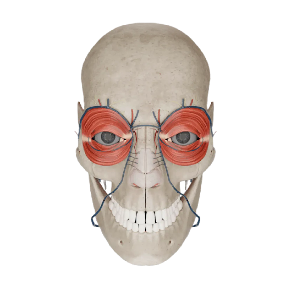
16. Which extraocular muscle is innervated by the trochlear nerve (IV cranial nerve)?
Superior rectus muscle.
The trochlear nerve (n. trochlearis) provides motor innervation to only one eye muscle — the superior oblique (m. obliquus superior).
Superior oblique muscle.
The trochlear nerve (n. trochlearis) provides motor innervation to only one eye muscle — the superior oblique (m. obliquus superior).
Inferior oblique muscle.
The trochlear nerve (n. trochlearis) provides motor innervation to only one eye muscle — the superior oblique (m. obliquus superior).
Lateral rectus muscle.
The trochlear nerve (n. trochlearis) provides motor innervation to only one eye muscle — the superior oblique (m. obliquus superior).
I find it difficult to answer
The trochlear nerve (n. trochlearis) provides motor innervation to only one eye muscle — the superior oblique (m. obliquus superior).
17. The labyrinthine artery (a. labyrinthi), supplying the inner ear, most often branches from:
Internal carotid artery
The labyrinthine artery (a. labyrinthi) most commonly is a branch of the anterior inferior cerebellar artery (AICA) or directly from the basilar artery.
Posterior communicating artery.
The labyrinthine artery (a. labyrinthi) most commonly is a branch of the anterior inferior cerebellar artery (AICA) or directly from the basilar artery.
Anterior inferior cerebellar or basilar artery.
The labyrinthine artery (a. labyrinthi) most commonly is a branch of the anterior inferior cerebellar artery (AICA) or directly from the basilar artery.
Occipital artery.
The labyrinthine artery (a. labyrinthi) most commonly is a branch of the anterior inferior cerebellar artery (AICA) or directly from the basilar artery.
I find it difficult to answer
The labyrinthine artery (a. labyrinthi) most commonly is a branch of the anterior inferior cerebellar artery (AICA) or directly from the basilar artery.
18. What anatomical structure connects the tympanic cavity (cavitas tympani) with the nasopharynx?
External acoustic meatus
The auditory tube (tuba auditiva) provides communication from the tympanic cavity to the nasopharynx, allowing for pressure equalization.
Round window.
The auditory tube (tuba auditiva) provides communication from the tympanic cavity to the nasopharynx, allowing for pressure equalization.
Auditory (Eustachian) tube.
The auditory tube (tuba auditiva) provides communication from the tympanic cavity to the nasopharynx, allowing for pressure equalization.
Oval window.
The auditory tube (tuba auditiva) provides communication from the tympanic cavity to the nasopharynx, allowing for pressure equalization.
I find it difficult to answer
The auditory tube (tuba auditiva) provides communication from the tympanic cavity to the nasopharynx, allowing for pressure equalization.
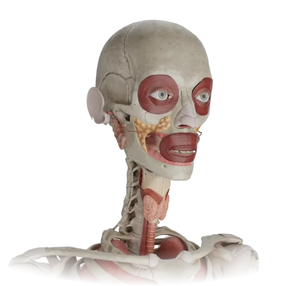
19. What important anatomical structures pass through the parenchyma of the parotid gland?
The facial nerve, forming a plexus, the retromandibular vein, and the external carotid artery pass through the parenchyma of the parotid gland (glandula parotidea).
The facial nerve, forming a plexus, the retromandibular vein, and the external carotid artery pass through the parenchyma of the parotid gland (glandula parotidea).
Hypoglossal nerve, lingual artery, lingual vein.
The facial nerve, forming a plexus, the retromandibular vein, and the external carotid artery pass through the parenchyma of the parotid gland (glandula parotidea).
The facial nerve, forming a plexus, the retromandibular vein, and the external carotid artery pass through the parenchyma of the parotid gland (glandula parotidea).
I find it difficult to answer
The facial nerve, forming a plexus, the retromandibular vein, and the external carotid artery pass through the parenchyma of the parotid gland (glandula parotidea).
20. Where does the parotid gland duct (ductus parotideus) open into the vestibule of the mouth?
On the mucous membrane of the cheek at the level of the second upper molar tooth
The duct of the parotid gland (Stensen's duct) pierces the buccinator muscle and opens on the papilla of the mucous membrane of the cheek at the level of the second upper molar.
Along the lingual fold
The duct of the parotid gland (Stensen's duct) pierces the buccinator muscle and opens on the papilla of the mucous membrane of the cheek at the level of the second upper molar.
On the sublingual caruncle
The duct of the parotid gland (Stensen's duct) pierces the buccinator muscle and opens on the papilla of the mucous membrane of the cheek at the level of the second upper molar.
In the region of the palatine tonsil
The duct of the parotid gland (Stensen's duct) pierces the buccinator muscle and opens on the papilla of the mucous membrane of the cheek at the level of the second upper molar.
I find it difficult to answer
The duct of the parotid gland (Stensen's duct) pierces the buccinator muscle and opens on the papilla of the mucous membrane of the cheek at the level of the second upper molar.