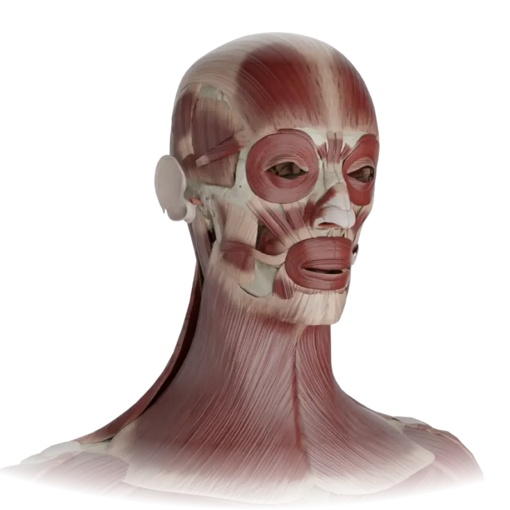
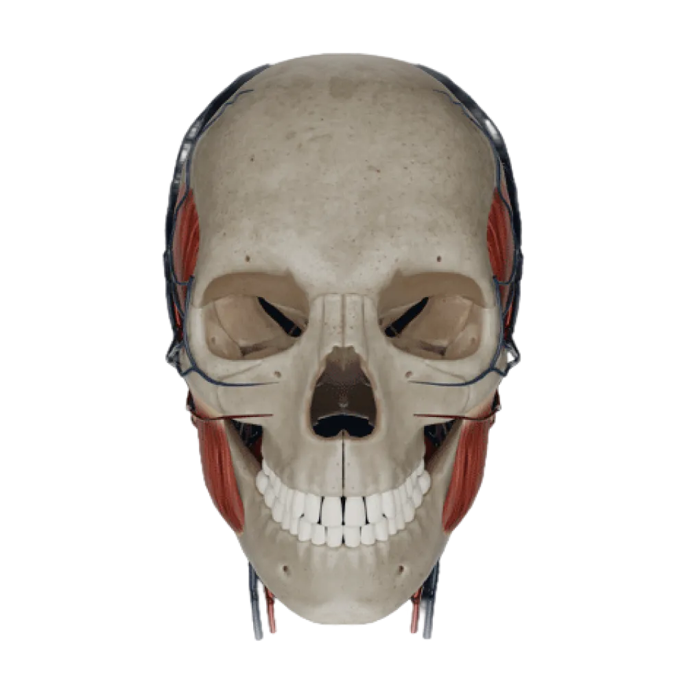
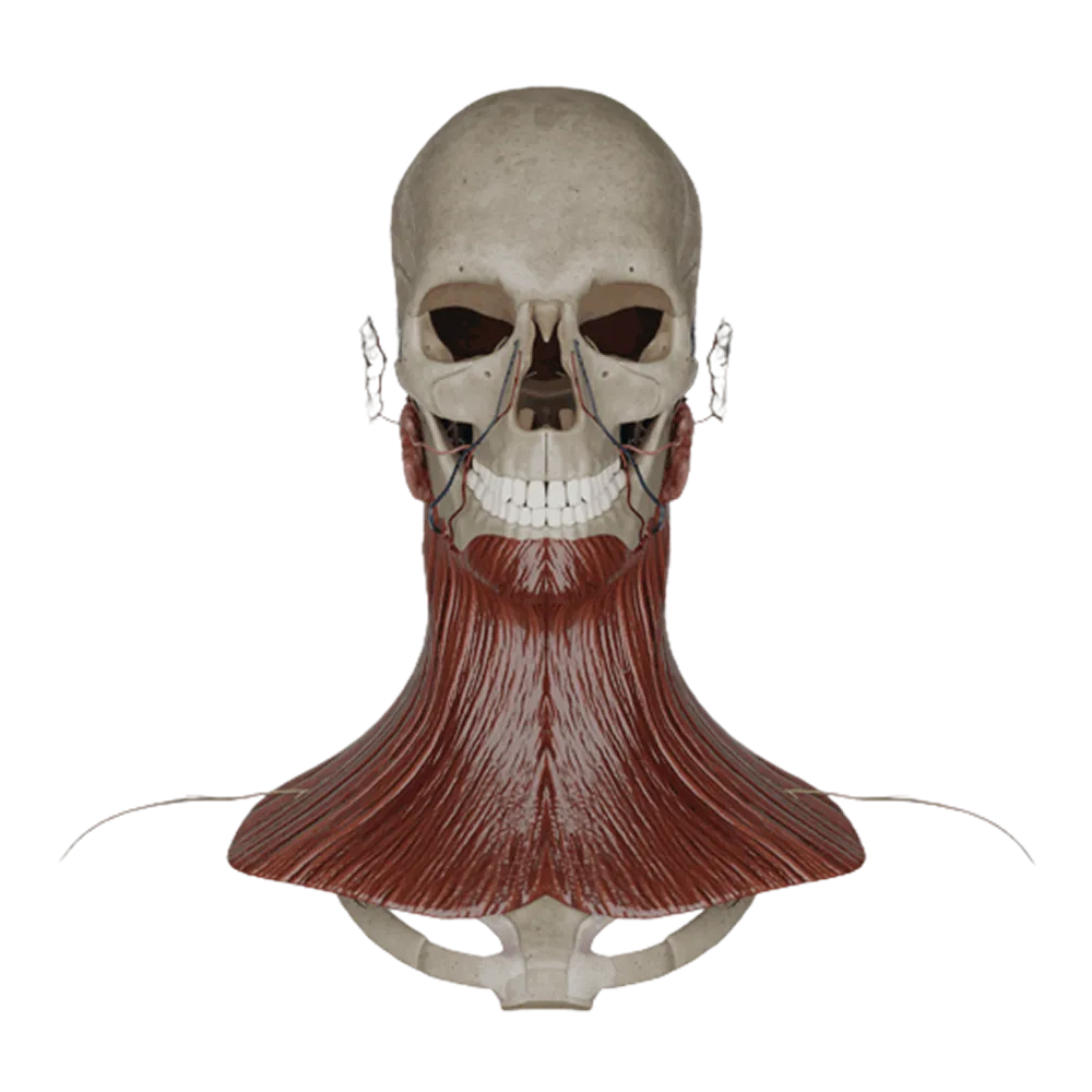
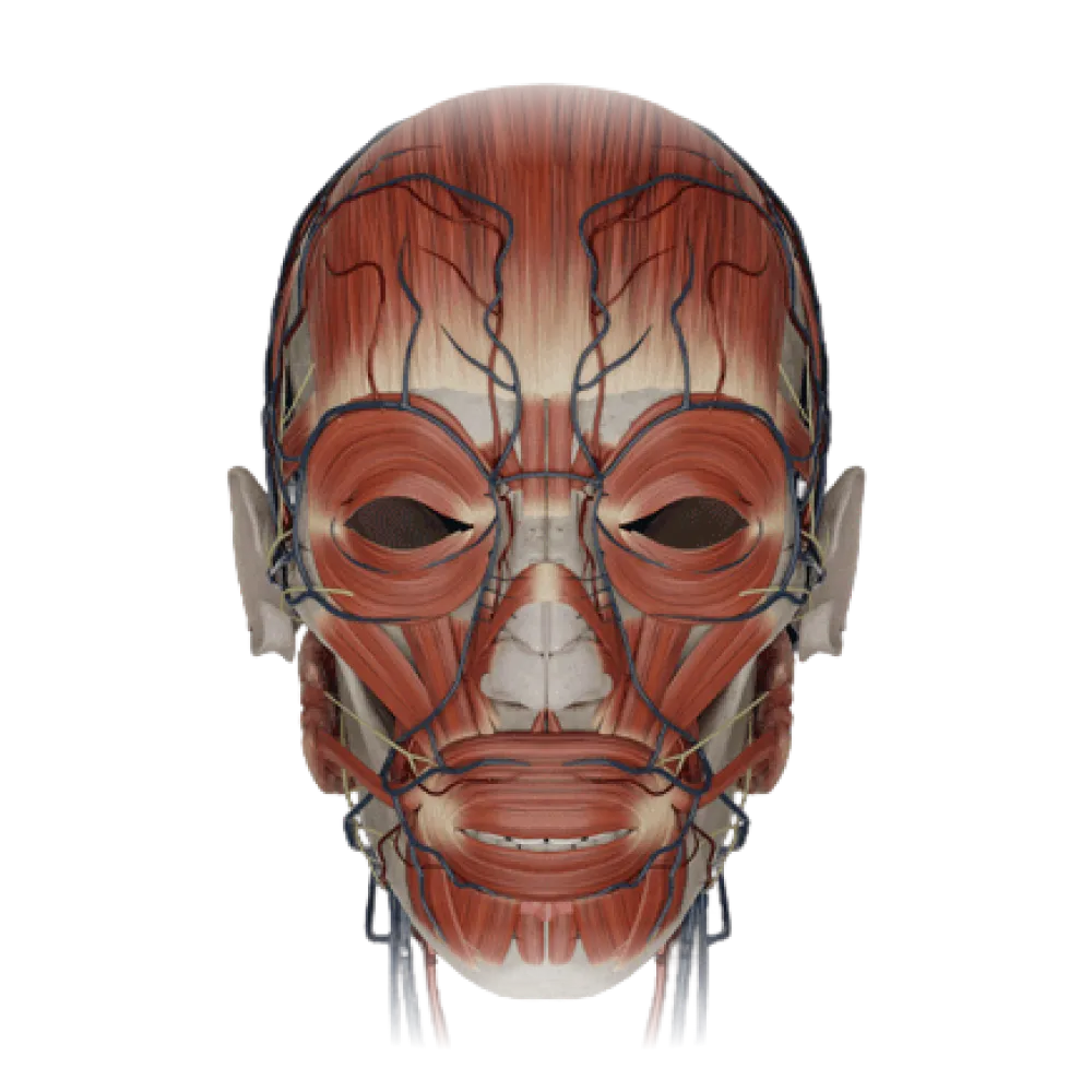
Anatomy test of the masticatory muscles
Evaluate the knowledge of the anatomy of the masticatory muscles. The test checks their topography, blood supply, venous drainage, and innervation.
1/20
bold
text
1. Which branch of the trigeminal nerve provides motor innervation to the masticatory muscles?
-
Ophthalmic nerve
The motor portion of the trigeminal nerve runs within its third branch, the mandibular nerve (n. mandibularis).
-
Maxillary nerve
The motor portion of the trigeminal nerve runs within its third branch, the mandibular nerve (n. mandibularis).
-
Mandibular nerve
The motor portion of the trigeminal nerve runs within its third branch, the mandibular nerve (n. mandibularis).
-
Facial nerve
The motor portion of the trigeminal nerve runs within its third branch, the mandibular nerve (n. mandibularis).
-
I find it difficult to answer
The motor portion of the trigeminal nerve runs within its third branch, the mandibular nerve (n. mandibularis).
2. Through which cranial foramen does the motor root of the trigeminal nerve exit the cranial cavity for innervation of the masticatory muscles?
-
Foramen rotundum
The mandibular nerve, containing motor fibers for the masticatory muscles, exits the cranial cavity through the oval foramen (foramen ovale).
-
Foramen spinosum
The mandibular nerve, containing motor fibers for the masticatory muscles, exits the cranial cavity through the oval foramen (foramen ovale).
-
Foramen lacerum
The mandibular nerve, containing motor fibers for the masticatory muscles, exits the cranial cavity through the oval foramen (foramen ovale).
-
Foramen ovale
The mandibular nerve, containing motor fibers for the masticatory muscles, exits the cranial cavity through the oval foramen (foramen ovale).
-
I find it difficult to answer
The mandibular nerve, containing motor fibers for the masticatory muscles, exits the cranial cavity through the oval foramen (foramen ovale).
3. Which artery is the main source of blood supply to the masseter muscle (m. masseter)?
-
Masseteric artery
The masseter muscle is predominantly supplied by the masseteric artery (a. masseterica), which is a branch of the maxillary artery.
-
Facial artery
The masseter muscle is predominantly supplied by the masseteric artery (a. masseterica), which is a branch of the maxillary artery.
-
Superficial temporal artery
The masseter muscle is predominantly supplied by the masseteric artery (a. masseterica), which is a branch of the maxillary artery.
-
Buccal artery
The masseter muscle is predominantly supplied by the masseteric artery (a. masseterica), which is a branch of the maxillary artery.
-
I find it difficult to answer
The masseter muscle is predominantly supplied by the masseteric artery (a. masseterica), which is a branch of the maxillary artery.
4. From which artery do the deep temporal arteries (aa. temporales profundae) mainly arise, supplying the temporalis muscle?
-
From the superficial temporal artery
The deep temporal arteries (anterior and posterior) branch from the pterygoid segment of the maxillary artery (a. maxillaris).
-
From the facial artery
The deep temporal arteries (anterior and posterior) branch from the pterygoid segment of the maxillary artery (a. maxillaris).
-
From the maxillary artery
The deep temporal arteries (anterior and posterior) branch from the pterygoid segment of the maxillary artery (a. maxillaris).
-
From the posterior auricular artery
The deep temporal arteries (anterior and posterior) branch from the pterygoid segment of the maxillary artery (a. maxillaris).
-
I find it difficult to answer
The deep temporal arteries (anterior and posterior) branch from the pterygoid segment of the maxillary artery (a. maxillaris).
5. Which nerve innervates the lateral pterygoid muscle?
-
N. buccalis
The lateral pterygoid muscle is innervated by the homonymous nerve (n. pterygoideus lateralis), originating from the anterior trunk of the mandibular nerve.
-
N. pterygoideus lateralis
The lateral pterygoid muscle is innervated by the homonymous nerve (n. pterygoideus lateralis), originating from the anterior trunk of the mandibular nerve.
-
N. massetericus
The lateral pterygoid muscle is innervated by the homonymous nerve (n. pterygoideus lateralis), originating from the anterior trunk of the mandibular nerve.
-
N. pterygoideus medialis
The lateral pterygoid muscle is innervated by the homonymous nerve (n. pterygoideus lateralis), originating from the anterior trunk of the mandibular nerve.
-
I find it difficult to answer
The lateral pterygoid muscle is innervated by the homonymous nerve (n. pterygoideus lateralis), originating from the anterior trunk of the mandibular nerve.
6. Through which anatomical structure do the masseteric nerve and artery (n. et a. masseterica) pass to the masseter muscle?
-
Pterygomaxillary fissure
The masseteric neurovascular bundle passes from the infratemporal fossa to the outer surface of the ramus of the mandible through the mandibular notch (incisura mandibulae).
-
Foramen ovale
The masseteric neurovascular bundle passes from the infratemporal fossa to the outer surface of the ramus of the mandible through the mandibular notch (incisura mandibulae).
-
Mandibular notch
The masseteric neurovascular bundle passes from the infratemporal fossa to the outer surface of the ramus of the mandible through the mandibular notch (incisura mandibulae).
-
Submandibular triangle
The masseteric neurovascular bundle passes from the infratemporal fossa to the outer surface of the ramus of the mandible through the mandibular notch (incisura mandibulae).
-
I find it difficult to answer
The masseteric neurovascular bundle passes from the infratemporal fossa to the outer surface of the ramus of the mandible through the mandibular notch (incisura mandibulae).
7. To which venous plexus does blood predominantly drain from the masticatory muscles?
-
To the cavernous sinus
Venous drainage from the masticatory muscles is primarily into the pterygoid venous plexus (plexus venosus pterygoideus), located in the infratemporal fossa.
-
To the facial vein
Venous drainage from the masticatory muscles is primarily into the pterygoid venous plexus (plexus venosus pterygoideus), located in the infratemporal fossa.
-
To the pharyngeal venous plexus
Venous drainage from the masticatory muscles is primarily into the pterygoid venous plexus (plexus venosus pterygoideus), located in the infratemporal fossa.
-
To the pterygoid venous plexus
Venous drainage from the masticatory muscles is primarily into the pterygoid venous plexus (plexus venosus pterygoideus), located in the infratemporal fossa.
-
I find it difficult to answer
Venous drainage from the masticatory muscles is primarily into the pterygoid venous plexus (plexus venosus pterygoideus), located in the infratemporal fossa.
8. The middle temporal artery (a. temporalis media), contributing to the blood supply of the temporalis muscle, branches from which artery?
-
Maxillary artery
The middle temporal artery arises from the superficial temporal artery (a. temporalis superficialis) and pierces the temporal fascia to supply the muscle.
-
Superficial temporal artery
The middle temporal artery arises from the superficial temporal artery (a. temporalis superficialis) and pierces the temporal fascia to supply the muscle.
-
Facial artery
The middle temporal artery arises from the superficial temporal artery (a. temporalis superficialis) and pierces the temporal fascia to supply the muscle.
-
Occipital artery.
The middle temporal artery arises from the superficial temporal artery (a. temporalis superficialis) and pierces the temporal fascia to supply the muscle.
-
I find it difficult to answer
The middle temporal artery arises from the superficial temporal artery (a. temporalis superficialis) and pierces the temporal fascia to supply the muscle.
9. Which muscle, derived from the first pharyngeal arch, is innervated by the mylohyoid nerve (n. mylohyoideus) along with the masticatory muscles?
-
Anterior belly of the digastric muscle.
The anterior belly of the digastric muscle (as well as the mylohyoid muscle) originates from the 1st pharyngeal arch and is innervated by the n. Mylohyoideus from CN V
-
Posterior belly of the digastric muscle.
The anterior belly of the digastric muscle (as well as the mylohyoid muscle) originates from the 1st pharyngeal arch and is innervated by the n. Mylohyoideus from CN V
-
Stylohyoid muscle
The anterior belly of the digastric muscle (as well as the mylohyoid muscle) originates from the 1st pharyngeal arch and is innervated by the n. Mylohyoideus from CN V
-
Submental muscle
The anterior belly of the digastric muscle (as well as the mylohyoid muscle) originates from the 1st pharyngeal arch and is innervated by the n. Mylohyoideus from CN V
-
I find it difficult to answer
The anterior belly of the digastric muscle (as well as the mylohyoid muscle) originates from the 1st pharyngeal arch and is innervated by the n. Mylohyoideus from CN V
10. From which division of the mandibular nerve does the medial pterygoid nerve (n. pterygoideus medialis) arise?
-
From the posterior trunk
The medial pterygoid nerve directly branches from the main trunk of the mandibular nerve on its medial side immediately after exiting the oval foramen.
-
From the auriculotemporal nerve
The medial pterygoid nerve directly branches from the main trunk of the mandibular nerve on its medial side immediately after exiting the oval foramen.
-
From the main trunk before its division
The medial pterygoid nerve directly branches from the main trunk of the mandibular nerve on its medial side immediately after exiting the oval foramen.
-
From the inferior alveolar nerve
The medial pterygoid nerve directly branches from the main trunk of the mandibular nerve on its medial side immediately after exiting the oval foramen.
-
I find it difficult to answer
The medial pterygoid nerve directly branches from the main trunk of the mandibular nerve on its medial side immediately after exiting the oval foramen.
11. Between which structures does the maxillary artery typically pass in the infratemporal fossa?
-
Between the medial and lateral pterygoid muscles
In the infratemporal fossa, the maxillary artery is most often found on the lateral surface of the lateral pterygoid muscle or between the lateral and medial pterygoid muscles.
-
Between the masseter and temporalis muscles
In the infratemporal fossa, the maxillary artery is most often found on the lateral surface of the lateral pterygoid muscle or between the lateral and medial pterygoid muscles.
-
Between the temporalis muscle and the cranial bones
In the infratemporal fossa, the maxillary artery is most often found on the lateral surface of the lateral pterygoid muscle or between the lateral and medial pterygoid muscles.
-
Between the styloid process and the digastric muscle
In the infratemporal fossa, the maxillary artery is most often found on the lateral surface of the lateral pterygoid muscle or between the lateral and medial pterygoid muscles.
-
I find it difficult to answer
In the infratemporal fossa, the maxillary artery is most often found on the lateral surface of the lateral pterygoid muscle or between the lateral and medial pterygoid muscles.
12. In addition to the masticatory muscles, which other muscle is innervated by motor fibers through n. Pterygoideus medialis
-
The muscle that elevates the soft palate
Tensor veli palatini (m. tensor veli palatini) is innervated by the homonymous nerve, branching out from the mandibular nerve (V3) Pterygoideus medialis
-
Stylopharyngeus muscle
Tensor veli palatini (m. tensor veli palatini) is innervated by the homonymous nerve, branching out from the mandibular nerve (V3) Pterygoideus medialis
-
Palatoglossus muscle.
Tensor veli palatini (m. tensor veli palatini) is innervated by the homonymous nerve, branching out from the mandibular nerve (V3) Pterygoideus medialis
-
The muscle that tenses the soft palate
Tensor veli palatini (m. tensor veli palatini) is innervated by the homonymous nerve, branching out from the mandibular nerve (V3) Pterygoideus medialis
-
I find it difficult to answer
Tensor veli palatini (m. tensor veli palatini) is innervated by the homonymous nerve, branching out from the mandibular nerve (V3) Pterygoideus medialis
13. Which artery, involved in the blood supply of the jaws, branches from the first part of the maxillary artery?
-
Buccal artery
The inferior alveolar artery (a. alveolaris inferior) arises from the first part of the maxillary artery.
-
Masseteric artery
The inferior alveolar artery (a. alveolaris inferior) arises from the first part of the maxillary artery.
-
Inferior alveolar artery
The inferior alveolar artery (a. alveolaris inferior) arises from the first part of the maxillary artery.
-
Descending palatine artery
The inferior alveolar artery (a. alveolaris inferior) arises from the first part of the maxillary artery.
-
I find it difficult to answer
The inferior alveolar artery (a. alveolaris inferior) arises from the first part of the maxillary artery.
14. Which nerves innervate the temporalis muscle (m. temporalis)?
-
Superficial temporal nerves
The temporalis muscle is innervated by the deep temporal nerves (nn. temporales profundi), which branch from the anterior trunk of the mandibular nerve.
-
Auriculotemporal nerve
The temporalis muscle is innervated by the deep temporal nerves (nn. temporales profundi), which branch from the anterior trunk of the mandibular nerve.
-
Zygomaticotemporal nerve
The temporalis muscle is innervated by the deep temporal nerves (nn. temporales profundi), which branch from the anterior trunk of the mandibular nerve.
-
Deep temporal nerves
The temporalis muscle is innervated by the deep temporal nerves (nn. temporales profundi), which branch from the anterior trunk of the mandibular nerve.
-
I find it difficult to answer
The temporalis muscle is innervated by the deep temporal nerves (nn. temporales profundi), which branch from the anterior trunk of the mandibular nerve.
15. Which nerve provides sensory innervation to the capsule of the temporomandibular joint?
-
Auriculotemporal nerve
The capsule of the TMJ is primarily innervated by the articular branches of the auriculotemporal nerve (n. auriculotemporalis) and the masseteric nerve.
-
Buccal nerve
The capsule of the TMJ is primarily innervated by the articular branches of the auriculotemporal nerve (n. auriculotemporalis) and the masseteric nerve.
-
Lingual nerve
The capsule of the TMJ is primarily innervated by the articular branches of the auriculotemporal nerve (n. auriculotemporalis) and the masseteric nerve.
-
Inferior alveolar nerve
The capsule of the TMJ is primarily innervated by the articular branches of the auriculotemporal nerve (n. auriculotemporalis) and the masseteric nerve.
-
I find it difficult to answer
The capsule of the TMJ is primarily innervated by the articular branches of the auriculotemporal nerve (n. auriculotemporalis) and the masseteric nerve.
16. The pterygoid branches (rami pterygoidei), supplying the pterygoid muscles, arise from:
-
Superficial temporal artery
The pterygoid branches originate from the pterygoid (second) division of the maxillary artery.
-
Facial artery
The pterygoid branches originate from the pterygoid (second) division of the maxillary artery.
-
Maxillary artery
The pterygoid branches originate from the pterygoid (second) division of the maxillary artery.
-
Ascending pharyngeal artery
The pterygoid branches originate from the pterygoid (second) division of the maxillary artery.
-
I find it difficult to answer
The pterygoid branches originate from the pterygoid (second) division of the maxillary artery.
17. In which anatomical space is the pterygoid venous plexus located?
-
Infratemporal fossa
The pterygoid venous plexus is located in the infratemporal fossa (fossa infratemporalis) around the lateral pterygoid muscle.
-
Pterygopalatine fossa
The pterygoid venous plexus is located in the infratemporal fossa (fossa infratemporalis) around the lateral pterygoid muscle.
-
Submandibular triangle
The pterygoid venous plexus is located in the infratemporal fossa (fossa infratemporalis) around the lateral pterygoid muscle.
-
Retropharyngeal space
The pterygoid venous plexus is located in the infratemporal fossa (fossa infratemporalis) around the lateral pterygoid muscle.
-
I find it difficult to answer
The pterygoid venous plexus is located in the infratemporal fossa (fossa infratemporalis) around the lateral pterygoid muscle.
18. Which of the listed nerves passing near the masticatory muscles contains primarily motor fibers?
-
N. buccalis
The masseteric nerve (n. massetericus) is motor, unlike the sensory buccal, auriculotemporal, and lingual nerves.
-
N. auriculotemporalis
The masseteric nerve (n. massetericus) is motor, unlike the sensory buccal, auriculotemporal, and lingual nerves.
-
N. massetericus
The masseteric nerve (n. massetericus) is motor, unlike the sensory buccal, auriculotemporal, and lingual nerves.
-
N. lingualis
The masseteric nerve (n. massetericus) is motor, unlike the sensory buccal, auriculotemporal, and lingual nerves.
-
I find it difficult to answer
The masseteric nerve (n. massetericus) is motor, unlike the sensory buccal, auriculotemporal, and lingual nerves.
19. How does the buccal nerve (n. buccalis) relate to the lateral pterygoid muscle?
-
Passes medially to the muscle
The buccal nerve (a branch of n. mandibularis) typically passes between the superior and inferior heads of the lateral pterygoid muscle.
-
Rounds the inferior edge of the muscle
The buccal nerve (a branch of n. mandibularis) typically passes between the superior and inferior heads of the lateral pterygoid muscle.
-
Passes more superficial to the muscle
The buccal nerve (a branch of n. mandibularis) typically passes between the superior and inferior heads of the lateral pterygoid muscle.
-
Passes between the two heads of the lateral pterygoid muscle
The buccal nerve (a branch of n. mandibularis) typically passes between the superior and inferior heads of the lateral pterygoid muscle.
-
I find it difficult to answer
The buccal nerve (a branch of n. mandibularis) typically passes between the superior and inferior heads of the lateral pterygoid muscle.
20. Which of the masticatory muscles has the most superficial location and is covered by a dense homonymous fascia?
-
With the masseter muscle
The masseter muscle (m. masseter) is located superficially on the ramus of the mandible and is covered by a dense masseteric fascia.
-
With the temporalis muscle
The masseter muscle (m. masseter) is located superficially on the ramus of the mandible and is covered by a dense masseteric fascia.
-
With the lateral pterygoid muscle
The masseter muscle (m. masseter) is located superficially on the ramus of the mandible and is covered by a dense masseteric fascia.
-
With the medial pterygoid muscle
The masseter muscle (m. masseter) is located superficially on the ramus of the mandible and is covered by a dense masseteric fascia.
-
I find it difficult to answer
The masseter muscle (m. masseter) is located superficially on the ramus of the mandible and is covered by a dense masseteric fascia.
Retake this quiz?
Your current progress will be reset.