1/20
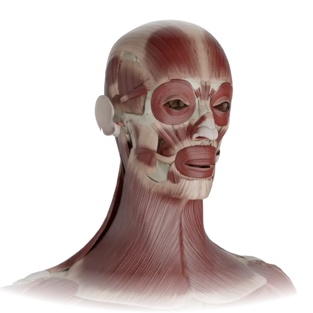
Test on the anatomy of the muscles of the head and neck.
Check your knowledge of the muscles of the head and neck. The test covers superficial, masticatory, and cervical muscles, their blood supply and innervation.
1. Which cranial nerve provides motor innervation to all mimetic muscles of the face?
-
Trigeminal nerve.
All mimetic muscles develop from the second branchial arch and are innervated by the facial nerve (n. facialis).
-
Facial nerve
All mimetic muscles develop from the second branchial arch and are innervated by the facial nerve (n. facialis).
-
Glossopharyngeal nerve
All mimetic muscles develop from the second branchial arch and are innervated by the facial nerve (n. facialis).
-
Vagus nerve
All mimetic muscles develop from the second branchial arch and are innervated by the facial nerve (n. facialis).
-
I find it difficult to answer
All mimetic muscles develop from the second branchial arch and are innervated by the facial nerve (n. facialis).
2. From which branchial arch do the masticatory muscles develop, which determines their innervation by the trigeminal nerve?
-
From the first branchial arch.
The masticatory muscles are derivatives of the first (mandibular) branchial arch and are innervated by the third branch of the trigeminal nerve (n. mandibularis).
-
From the second branchial arch.
The masticatory muscles are derivatives of the first (mandibular) branchial arch and are innervated by the third branch of the trigeminal nerve (n. mandibularis).
-
From the third branchial arch.
The masticatory muscles are derivatives of the first (mandibular) branchial arch and are innervated by the third branch of the trigeminal nerve (n. mandibularis).
-
From the fourth branchial arch.
The masticatory muscles are derivatives of the first (mandibular) branchial arch and are innervated by the third branch of the trigeminal nerve (n. mandibularis).
-
I find it difficult to answer
The masticatory muscles are derivatives of the first (mandibular) branchial arch and are innervated by the third branch of the trigeminal nerve (n. mandibularis).
3. What movement occurs with the unilateral contraction of the sternocleidomastoid muscle (m. sternocleidomastoideus)?
-
Tilting the head toward its side and turning the face in the opposite direction.
With unilateral contraction of m. sternocleidomastoideus tilts the head toward its side, and the face turns in the opposite direction.
-
Tilting the head and turning the face toward its side.
With unilateral contraction of m. sternocleidomastoideus tilts the head toward its side, and the face turns in the opposite direction.
-
Throwing the head back.
With unilateral contraction of m. sternocleidomastoideus tilts the head toward its side, and the face turns in the opposite direction.
-
Flexion of the cervical spine.
With unilateral contraction of m. sternocleidomastoideus tilts the head toward its side, and the face turns in the opposite direction.
-
I find it difficult to answer
With unilateral contraction of m. sternocleidomastoideus tilts the head toward its side, and the face turns in the opposite direction.
4. Which nerves innervate the anterior and posterior bellies of the digastric muscle (m. digastricus) respectively?
-
Trigeminal and facial nerves.
The anterior belly develops from the 1st arch (innervated by n. trigeminus), the posterior from the 2nd arch (innervated by n. facialis).
-
Facial and glossopharyngeal nerves.
The anterior belly develops from the 1st arch (innervated by n. trigeminus), the posterior from the 2nd arch (innervated by n. facialis).
-
Vagus and accessory nerves.
The anterior belly develops from the 1st arch (innervated by n. trigeminus), the posterior from the 2nd arch (innervated by n. facialis).
-
Trigeminal and hypoglossal nerves.
The anterior belly develops from the 1st arch (innervated by n. trigeminus), the posterior from the 2nd arch (innervated by n. facialis).
-
I find it difficult to answer
The anterior belly develops from the 1st arch (innervated by n. trigeminus), the posterior from the 2nd arch (innervated by n. facialis).
5. Which of the listed muscles, upon bilateral contraction, lowers the mandible (opens the mouth)?
-
Masseter muscle
Lateral pterygoid muscle (m. pterygoideus lateralis) upon bilateral contraction pulls the mandible forward and lowers it.
-
Temporalis muscle
Lateral pterygoid muscle (m. pterygoideus lateralis) upon bilateral contraction pulls the mandible forward and lowers it.
-
Medial pterygoid muscle
Lateral pterygoid muscle (m. pterygoideus lateralis) upon bilateral contraction pulls the mandible forward and lowers it.
-
Lateral pterygoid muscle
Lateral pterygoid muscle (m. pterygoideus lateralis) upon bilateral contraction pulls the mandible forward and lowers it.
-
I find it difficult to answer
Lateral pterygoid muscle (m. pterygoideus lateralis) upon bilateral contraction pulls the mandible forward and lowers it.
6. Which of the listed muscles does NOT belong to the infrahyoid muscles of the neck (musculi infrahyoidei)?
-
Sternohyoid muscle.
Mylohyoid muscle (m. mylohyoideus) is located above the hyoid bone and belongs to the suprahyoid muscles.
-
Omohyoid muscle
Mylohyoid muscle (m. mylohyoideus) is located above the hyoid bone and belongs to the suprahyoid muscles.
-
Mylohyoid muscle
Mylohyoid muscle (m. mylohyoideus) is located above the hyoid bone and belongs to the suprahyoid muscles.
-
Thyrohyoid muscle.
Mylohyoid muscle (m. mylohyoideus) is located above the hyoid bone and belongs to the suprahyoid muscles.
-
I find it difficult to answer
Mylohyoid muscle (m. mylohyoideus) is located above the hyoid bone and belongs to the suprahyoid muscles.
7. Which fascia covers the buccinator muscle (m. buccinator) and transitions to the superior pharyngeal constrictor?
-
Masseteric fascia.
The buccopharyngeal fascia (fascia buccopharyngea) covers the buccinator muscle and extends posteriorly to the lateral wall of the pharynx.
-
Temporal fascia.
The buccopharyngeal fascia (fascia buccopharyngea) covers the buccinator muscle and extends posteriorly to the lateral wall of the pharynx.
-
Buccopharyngeal fascia.
The buccopharyngeal fascia (fascia buccopharyngea) covers the buccinator muscle and extends posteriorly to the lateral wall of the pharynx.
-
Superficial fascia of the face.
The buccopharyngeal fascia (fascia buccopharyngea) covers the buccinator muscle and extends posteriorly to the lateral wall of the pharynx.
-
I find it difficult to answer
The buccopharyngeal fascia (fascia buccopharyngea) covers the buccinator muscle and extends posteriorly to the lateral wall of the pharynx.
8. What is the name of the tendinous helmet that connects the frontal and occipital bellies of the epicranial muscle (m. epicranius)?
-
Galea aponeurotica.
The tendinous helmet (galea aponeurotica) is tightly fused with the skin of the scalp and loosely with the periosteum of the bones of the skullcap.
-
Fascia temporalis.
The tendinous helmet (galea aponeurotica) is tightly fused with the skin of the scalp and loosely with the periosteum of the bones of the skullcap.
-
Aponeurosis palatina.
The tendinous helmet (galea aponeurotica) is tightly fused with the skin of the scalp and loosely with the periosteum of the bones of the skullcap.
-
Fascia masseterica.
The tendinous helmet (galea aponeurotica) is tightly fused with the skin of the scalp and loosely with the periosteum of the bones of the skullcap.
-
I find it difficult to answer
The tendinous helmet (galea aponeurotica) is tightly fused with the skin of the scalp and loosely with the periosteum of the bones of the skullcap.
9. In which layer (according to V.N. Shevkunenko's classification of the fascial layers of the neck) is the platysma located?
-
In the separation of the superficial fascia (first fascia).
Platysma is located in the subcutaneous tissue and is closely associated with the superficial fascia of the neck (fascia colli superficialis).
-
Under the superficial layer of the investing fascia (second fascia).
Platysma is located in the subcutaneous tissue and is closely associated with the superficial fascia of the neck (fascia colli superficialis).
-
Within the omoclavicular fascia (third fascia).
Platysma is located in the subcutaneous tissue and is closely associated with the superficial fascia of the neck (fascia colli superficialis).
-
Inside the endocervical fascia (fourth fascia).
Platysma is located in the subcutaneous tissue and is closely associated with the superficial fascia of the neck (fascia colli superficialis).
-
I find it difficult to answer
Platysma is located in the subcutaneous tissue and is closely associated with the superficial fascia of the neck (fascia colli superficialis).
10. Between which neck muscles is the interscalenic space (spatium interscalenum) located, where the subclavian artery and the brachial plexus pass?
-
Between the sternocleidomastoid and anterior scalene muscles.
The interscalenic space is bordered anteriorly by m. scalenus anterior, and posteriorly by m. scalenus medius, and below by the first rib.
-
Between the anterior and middle scalenes.
The interscalenic space is bordered anteriorly by m. scalenus anterior, and posteriorly by m. scalenus medius, and below by the first rib.
-
Between the middle and posterior scalene muscles.
The interscalenic space is bordered anteriorly by m. scalenus anterior, and posteriorly by m. scalenus medius, and below by the first rib.
-
Between the longus colli and the anterior scalene muscle.
The interscalenic space is bordered anteriorly by m. scalenus anterior, and posteriorly by m. scalenus medius, and below by the first rib.
-
I find it difficult to answer
The interscalenic space is bordered anteriorly by m. scalenus anterior, and posteriorly by m. scalenus medius, and below by the first rib.
11. Which muscle forms the muscular floor of the oral diaphragm (diaphragma oris)?
-
Digastric muscle
The mylohyoid muscle (m. mylohyoideus) is paired, merging at the midline to form the floor (diaphragm) of the oral cavity.
-
Mylohyoid muscle
The mylohyoid muscle (m. mylohyoideus) is paired, merging at the midline to form the floor (diaphragm) of the oral cavity.
-
Geniohyoid muscle
The mylohyoid muscle (m. mylohyoideus) is paired, merging at the midline to form the floor (diaphragm) of the oral cavity.
-
Stylohyoid muscle
The mylohyoid muscle (m. mylohyoideus) is paired, merging at the midline to form the floor (diaphragm) of the oral cavity.
-
I find it difficult to answer
The mylohyoid muscle (m. mylohyoideus) is paired, merging at the midline to form the floor (diaphragm) of the oral cavity.
12. To which anatomical structure of the mandible does the temporal muscle (m. temporalis) attach?
-
Coronoid process.
The powerful tendon of the temporal muscle attaches to the coronoid process (processus coronoideus) of the mandible.
-
Condyle process.
The powerful tendon of the temporal muscle attaches to the coronoid process (processus coronoideus) of the mandible.
-
Neck of the mandible.
The powerful tendon of the temporal muscle attaches to the coronoid process (processus coronoideus) of the mandible.
-
Angle of the mandible.
The powerful tendon of the temporal muscle attaches to the coronoid process (processus coronoideus) of the mandible.
-
I find it difficult to answer
The powerful tendon of the temporal muscle attaches to the coronoid process (processus coronoideus) of the mandible.
13. Which muscle is part of the deep muscles of the neck (prevertebral group)?
-
Splenius colli muscle.
The longus colli muscle (m. longus colli) is located on the anterior surface of the vertebral bodies from the atlas to the 3rd thoracic vertebra and belongs to the prevertebral group.
-
Longus colli muscle.
The longus colli muscle (m. longus colli) is located on the anterior surface of the vertebral bodies from the atlas to the 3rd thoracic vertebra and belongs to the prevertebral group.
-
Levator scapulae.
The longus colli muscle (m. longus colli) is located on the anterior surface of the vertebral bodies from the atlas to the 3rd thoracic vertebra and belongs to the prevertebral group.
-
Semispinalis capitis muscle
The longus colli muscle (m. longus colli) is located on the anterior surface of the vertebral bodies from the atlas to the 3rd thoracic vertebra and belongs to the prevertebral group.
-
I find it difficult to answer
The longus colli muscle (m. longus colli) is located on the anterior surface of the vertebral bodies from the atlas to the 3rd thoracic vertebra and belongs to the prevertebral group.
14. Which cranial nerve provides motor innervation to the sternocleidomastoid and trapezius muscles?
-
Glossopharyngeal nerve
Accessory nerve (n. accessorius, XI pair) innervates m. sternocleidomastoideus and m. trapezius.
-
Vagus nerve
Accessory nerve (n. accessorius, XI pair) innervates m. sternocleidomastoideus and m. trapezius.
-
Accessory nerve
Accessory nerve (n. accessorius, XI pair) innervates m. sternocleidomastoideus and m. trapezius.
-
Hypoglossal nerve
Accessory nerve (n. accessorius, XI pair) innervates m. sternocleidomastoideus and m. trapezius.
-
I find it difficult to answer
Accessory nerve (n. accessorius, XI pair) innervates m. sternocleidomastoideus and m. trapezius.
15. Which muscle closes the eyelid aperture when contracted?
-
Orbicularis oculi muscle.
The orbicularis oculi muscle (m. orbicularis oculi) consists of orbital, palpebral, and lacrimal parts; its main function is closing the eyelids.
-
Corrugator supercilii muscle.
The orbicularis oculi muscle (m. orbicularis oculi) consists of orbital, palpebral, and lacrimal parts; its main function is closing the eyelids.
-
Procerus muscle.
The orbicularis oculi muscle (m. orbicularis oculi) consists of orbital, palpebral, and lacrimal parts; its main function is closing the eyelids.
-
Frontal belly of the epicranial muscle.
The orbicularis oculi muscle (m. orbicularis oculi) consists of orbital, palpebral, and lacrimal parts; its main function is closing the eyelids.
-
I find it difficult to answer
The orbicularis oculi muscle (m. orbicularis oculi) consists of orbital, palpebral, and lacrimal parts; its main function is closing the eyelids.
16. From which bony structure does the masseter muscle (m. masseter) originate?
-
From the temporal fossa.
The masseter muscle originates from the lower margin and inner surface of the zygomatic arch and attaches to the masseteric tuberosity of the mandible.
-
From the zygomatic arch.
The masseter muscle originates from the lower margin and inner surface of the zygomatic arch and attaches to the masseteric tuberosity of the mandible.
-
From the pterygoid process of the sphenoid bone.
The masseter muscle originates from the lower margin and inner surface of the zygomatic arch and attaches to the masseteric tuberosity of the mandible.
-
From the mastoid process of the temporal bone.
The masseter muscle originates from the lower margin and inner surface of the zygomatic arch and attaches to the masseteric tuberosity of the mandible.
-
I find it difficult to answer
The masseter muscle originates from the lower margin and inner surface of the zygomatic arch and attaches to the masseteric tuberosity of the mandible.
17. Which muscle forms the posterior boundary of the sublingual triangle (Pirogov's triangle)?
-
Anterior belly of the digastric muscle.
Pirogov's triangle is bordered anteriorly by the free edge m. mylohyoideus, posteriorly by the posterior belly of m. digastrici, and superiorly by n. hypoglossus.
-
Mylohyoid muscle
Pirogov's triangle is bordered anteriorly by the free edge m. mylohyoideus, posteriorly by the posterior belly of m. digastrici, and superiorly by n. hypoglossus.
-
Posterior belly of the digastric muscle.
Pirogov's triangle is bordered anteriorly by the free edge m. mylohyoideus, posteriorly by the posterior belly of m. digastrici, and superiorly by n. hypoglossus.
-
Hyoglossus muscle.
Pirogov's triangle is bordered anteriorly by the free edge m. mylohyoideus, posteriorly by the posterior belly of m. digastrici, and superiorly by n. hypoglossus.
-
I find it difficult to answer
Pirogov's triangle is bordered anteriorly by the free edge m. mylohyoideus, posteriorly by the posterior belly of m. digastrici, and superiorly by n. hypoglossus.
18. Which neck muscles are covered by the omoclavicular fascia (the third fascia of the neck according to Shevkunenko)?
-
Muscles lying above the hyoid bone.
The third fascia of the neck (fascia omoclavicularis) forms sheaths for infrahyoid muscles of the neck.
-
Muscles lying below the hyoid bone.
The third fascia of the neck (fascia omoclavicularis) forms sheaths for infrahyoid muscles of the neck.
-
The deep scalene muscles.
The third fascia of the neck (fascia omoclavicularis) forms sheaths for infrahyoid muscles of the neck.
-
Prevertebral muscles.
The third fascia of the neck (fascia omoclavicularis) forms sheaths for infrahyoid muscles of the neck.
-
I find it difficult to answer
The third fascia of the neck (fascia omoclavicularis) forms sheaths for infrahyoid muscles of the neck.
19. Which mimic muscle pulls the corner of the mouth laterally and forms a dimple on the cheek?
-
Zygomaticus major muscle
The risorius muscle (m. risorius) is an inconsistent muscle that pulls the corner of the mouth laterally.
-
Risorius muscle
The risorius muscle (m. risorius) is an inconsistent muscle that pulls the corner of the mouth laterally.
-
Depressor anguli oris muscle
The risorius muscle (m. risorius) is an inconsistent muscle that pulls the corner of the mouth laterally.
-
Buccinator
The risorius muscle (m. risorius) is an inconsistent muscle that pulls the corner of the mouth laterally.
-
I find it difficult to answer
The risorius muscle (m. risorius) is an inconsistent muscle that pulls the corner of the mouth laterally.
20. Which muscle divides the lateral region of the neck into the omotrapezoid and omoclavicular triangles?
-
Sternocleidomastoid muscle
The inferior belly of the omohyoid muscle (venter inferior m. omohyoidei) passes through the lateral region of the neck, dividing it into the mentioned triangles.
-
Omohyoid muscle
The inferior belly of the omohyoid muscle (venter inferior m. omohyoidei) passes through the lateral region of the neck, dividing it into the mentioned triangles.
-
Anterior scalene muscle
The inferior belly of the omohyoid muscle (venter inferior m. omohyoidei) passes through the lateral region of the neck, dividing it into the mentioned triangles.
-
Digastric muscle
The inferior belly of the omohyoid muscle (venter inferior m. omohyoidei) passes through the lateral region of the neck, dividing it into the mentioned triangles.
-
I find it difficult to answer
The inferior belly of the omohyoid muscle (venter inferior m. omohyoidei) passes through the lateral region of the neck, dividing it into the mentioned triangles.
Retake this quiz?
Your current progress will be reset.
Muscles of the head and neck
Superficial muscles of head
0/20