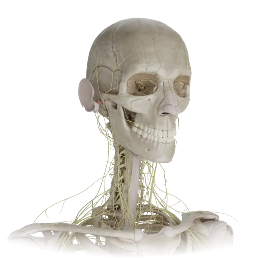
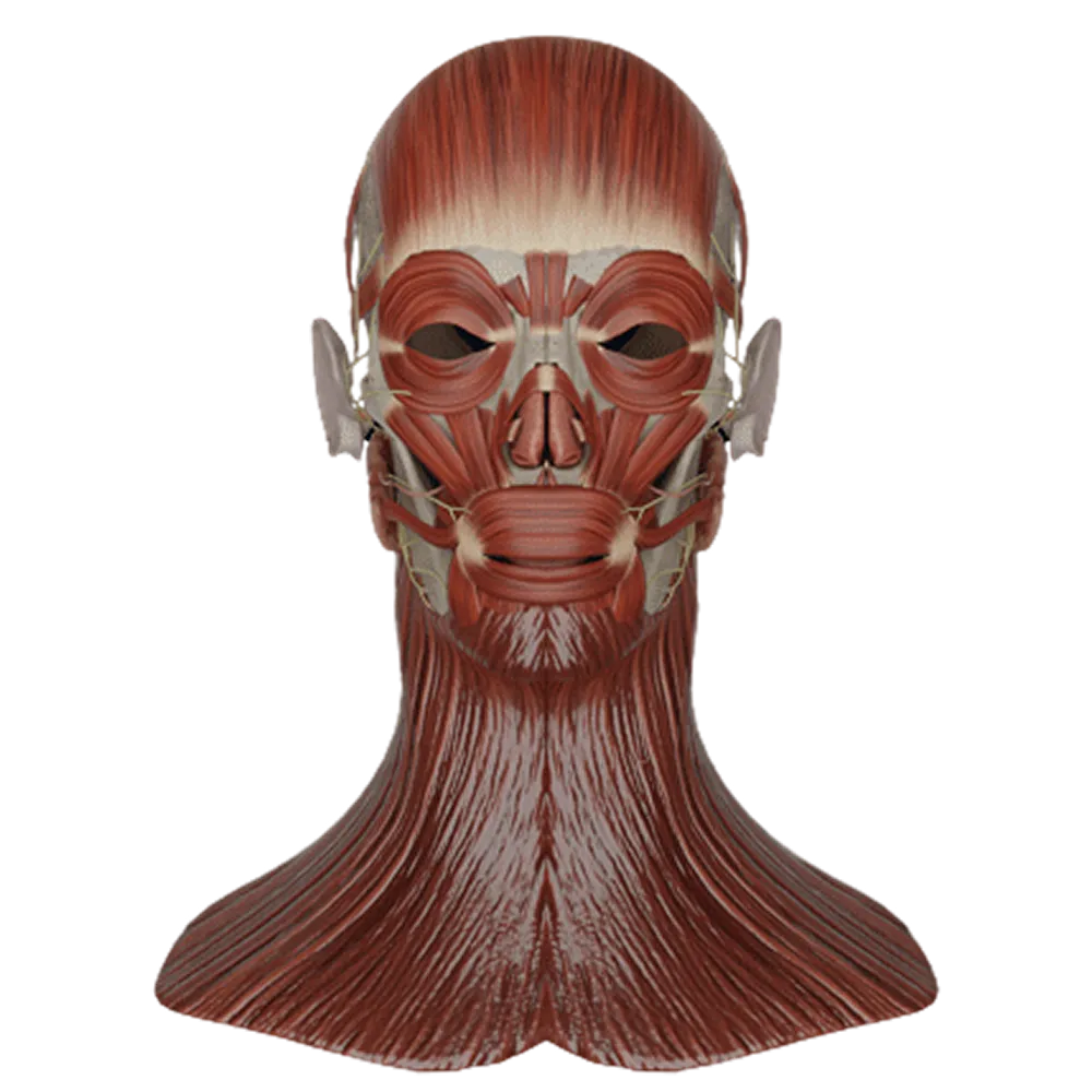
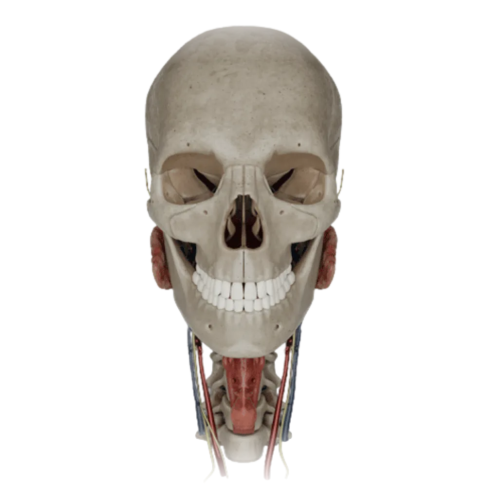
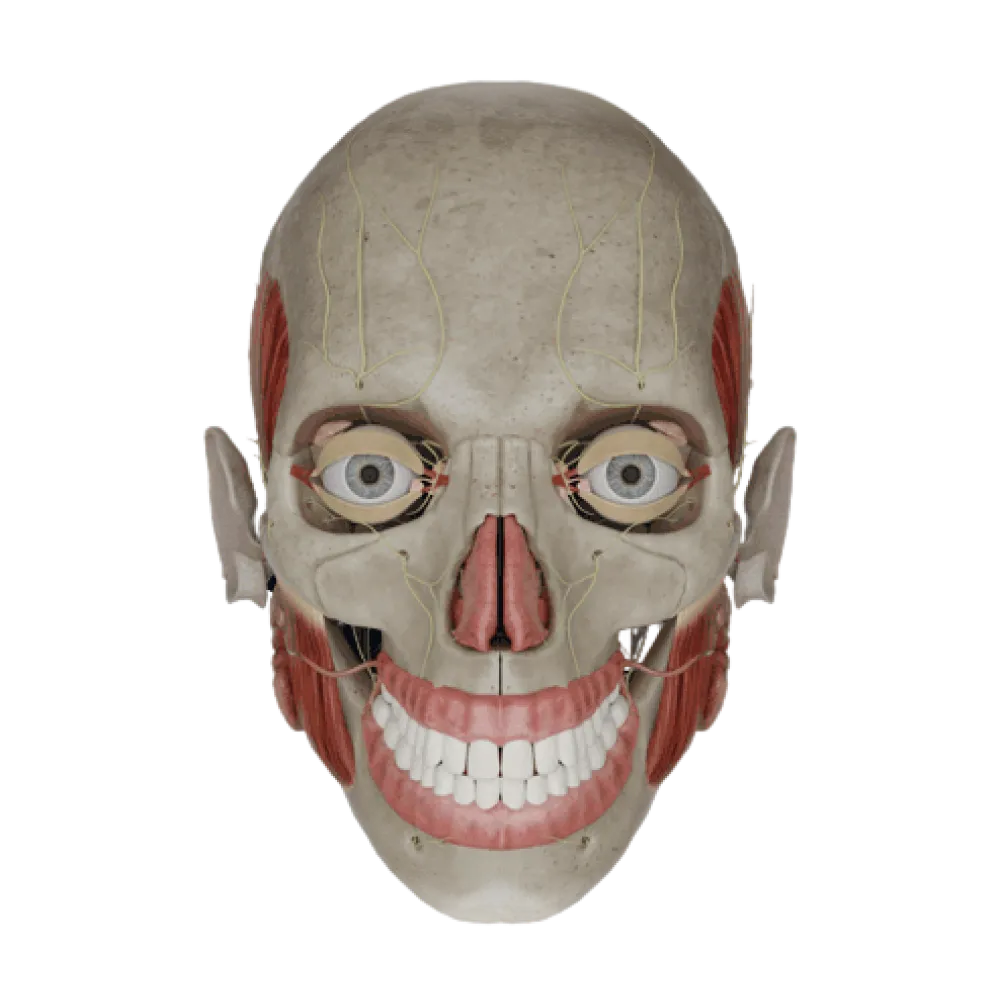
Facial nerve anatomy test
Evaluate the knowledge of the facial nerve anatomy. The test examines the nuclei, topography, branches, and also the motor, autonomic, and sensory zones.
1/20
bold
text
1. Where is the motor nucleus of the facial nerve located?
-
In the medulla oblongata
The motor nucleus of the facial nerve (nucleus n. facialis) lies in the reticular formation of the pontine tegmentum.
-
In the cerebral peduncles
The motor nucleus of the facial nerve (nucleus n. facialis) lies in the reticular formation of the pontine tegmentum.
-
In the roof of the midbrain
The motor nucleus of the facial nerve (nucleus n. facialis) lies in the reticular formation of the pontine tegmentum.
-
In the pontine tegmentum
The motor nucleus of the facial nerve (nucleus n. facialis) lies in the reticular formation of the pontine tegmentum.
-
I find it difficult to answer
The motor nucleus of the facial nerve (nucleus n. facialis) lies in the reticular formation of the pontine tegmentum.
2. Through which foramen does the facial nerve exit the skull?
-
Foramen rotundum
The facial nerve exits the facial canal of the temporal bone through the stylomastoid foramen (foramen stylomastoideum).
-
Foramen ovale
The facial nerve exits the facial canal of the temporal bone through the stylomastoid foramen (foramen stylomastoideum).
-
Stylomastoid foramen
The facial nerve exits the facial canal of the temporal bone through the stylomastoid foramen (foramen stylomastoideum).
-
Foramen lacerum
The facial nerve exits the facial canal of the temporal bone through the stylomastoid foramen (foramen stylomastoideum).
-
I find it difficult to answer
The facial nerve exits the facial canal of the temporal bone through the stylomastoid foramen (foramen stylomastoideum).
3. Which branch of the facial nerve branches off in the facial canal and innervates the lacrimal gland?
-
Greater petrosal nerve
The greater petrosal nerve (n. petrosus major) carries parasympathetic fibers to the pterygopalatine ganglion for the innervation of the lacrimal gland.
-
Chorda tympani
The greater petrosal nerve (n. petrosus major) carries parasympathetic fibers to the pterygopalatine ganglion for the innervation of the lacrimal gland.
-
Lesser petrosal nerve
The greater petrosal nerve (n. petrosus major) carries parasympathetic fibers to the pterygopalatine ganglion for the innervation of the lacrimal gland.
-
Stapedial nerve
The greater petrosal nerve (n. petrosus major) carries parasympathetic fibers to the pterygopalatine ganglion for the innervation of the lacrimal gland.
-
I find it difficult to answer
The greater petrosal nerve (n. petrosus major) carries parasympathetic fibers to the pterygopalatine ganglion for the innervation of the lacrimal gland.
4. What fibers does the chorda tympani contain?
-
Only motor
The chorda tympani contains special sensory (gustatory) fibers for the anterior two-thirds of the tongue and preganglionic parasympathetic fibers for the submandibular ganglion.
-
Only sympathetic
The chorda tympani contains special sensory (gustatory) fibers for the anterior two-thirds of the tongue and preganglionic parasympathetic fibers for the submandibular ganglion.
-
Gustatory and parasympathetic
The chorda tympani contains special sensory (gustatory) fibers for the anterior two-thirds of the tongue and preganglionic parasympathetic fibers for the submandibular ganglion.
-
General sensory and motor
The chorda tympani contains special sensory (gustatory) fibers for the anterior two-thirds of the tongue and preganglionic parasympathetic fibers for the submandibular ganglion.
-
I find it difficult to answer
The chorda tympani contains special sensory (gustatory) fibers for the anterior two-thirds of the tongue and preganglionic parasympathetic fibers for the submandibular ganglion.
5. Which muscle is innervated by the cervical branch (r. colli) of the facial nerve?
-
Sternocleidomastoid muscle
The cervical branch descends on the neck and innervates the platysma muscle, forming the superficial cervical loop with the transverse cervical nerve.
-
Platysma
The cervical branch descends on the neck and innervates the platysma muscle, forming the superficial cervical loop with the transverse cervical nerve.
-
Mylohyoid muscle
The cervical branch descends on the neck and innervates the platysma muscle, forming the superficial cervical loop with the transverse cervical nerve.
-
Digastric muscle
The cervical branch descends on the neck and innervates the platysma muscle, forming the superficial cervical loop with the transverse cervical nerve.
-
I find it difficult to answer
The cervical branch descends on the neck and innervates the platysma muscle, forming the superficial cervical loop with the transverse cervical nerve.
6. Where is the geniculate ganglion located?
-
In the pterygopalatine fossa
The geniculate ganglion is located at the bend of the facial canal in the temporal bone and contains pseudounipolar sensory neurons of the intermediate nerve.
-
In the bend of the facial nerve canal
The geniculate ganglion is located at the bend of the facial canal in the temporal bone and contains pseudounipolar sensory neurons of the intermediate nerve.
-
In the cerebellopontine angle
The geniculate ganglion is located at the bend of the facial canal in the temporal bone and contains pseudounipolar sensory neurons of the intermediate nerve.
-
In the parotid gland
The geniculate ganglion is located at the bend of the facial canal in the temporal bone and contains pseudounipolar sensory neurons of the intermediate nerve.
-
I find it difficult to answer
The geniculate ganglion is located at the bend of the facial canal in the temporal bone and contains pseudounipolar sensory neurons of the intermediate nerve.
7. To which muscle does the stapedial nerve (n. stapedius) lead?
-
To the stapedius muscle
The stapedial nerve is a motor branch of the facial nerve, branching off in the facial canal, and innervates the stapedius muscle in the tympanic cavity.
-
To the tensor tympani muscle
The stapedial nerve is a motor branch of the facial nerve, branching off in the facial canal, and innervates the stapedius muscle in the tympanic cavity.
-
To the levator veli palatini muscle
The stapedial nerve is a motor branch of the facial nerve, branching off in the facial canal, and innervates the stapedius muscle in the tympanic cavity.
-
To the styloglossus muscle
The stapedial nerve is a motor branch of the facial nerve, branching off in the facial canal, and innervates the stapedius muscle in the tympanic cavity.
-
I find it difficult to answer
The stapedial nerve is a motor branch of the facial nerve, branching off in the facial canal, and innervates the stapedius muscle in the tympanic cavity.
8. Which facial muscles are innervated by the buccal branches of the facial nerve?
-
Frontal belly of the occipitofrontalis muscle
The buccal branches (rr. buccales) innervate the buccinator muscle, orbicularis oris muscle, risorius, and muscles that elevate the upper lip and corner of the mouth.
-
Orbicularis oculi muscle.
The buccal branches (rr. buccales) innervate the buccinator muscle, orbicularis oris muscle, risorius, and muscles that elevate the upper lip and corner of the mouth.
-
Platysma
The buccal branches (rr. buccales) innervate the buccinator muscle, orbicularis oris muscle, risorius, and muscles that elevate the upper lip and corner of the mouth.
-
Buccinator and orbicularis oris muscle
The buccal branches (rr. buccales) innervate the buccinator muscle, orbicularis oris muscle, risorius, and muscles that elevate the upper lip and corner of the mouth.
-
I find it difficult to answer
The buccal branches (rr. buccales) innervate the buccinator muscle, orbicularis oris muscle, risorius, and muscles that elevate the upper lip and corner of the mouth.
9. With which nerve does the chorda tympani connect after leaving the tympanic cavity?
-
With the auriculotemporal nerve
After exiting the skull through the petrotympanic fissure, the chorda tympani joins the lingual nerve (a branch of the mandibular nerve).
-
With the lingual nerve
After exiting the skull through the petrotympanic fissure, the chorda tympani joins the lingual nerve (a branch of the mandibular nerve).
-
With the glossopharyngeal nerve
After exiting the skull through the petrotympanic fissure, the chorda tympani joins the lingual nerve (a branch of the mandibular nerve).
-
With the inferior alveolar nerve
After exiting the skull through the petrotympanic fissure, the chorda tympani joins the lingual nerve (a branch of the mandibular nerve).
-
I find it difficult to answer
After exiting the skull through the petrotympanic fissure, the chorda tympani joins the lingual nerve (a branch of the mandibular nerve).
10. Which nerve is the posterior auricular nerve (n. auricularis posterior) a branch of?
-
Trigeminal nerve.
The posterior auricular nerve branches from the facial nerve immediately after it exits the stylomastoid foramen and innervates the occipital belly of the occipitofrontalis muscle and the posterior auricular muscle.
-
Vagus nerve.
The posterior auricular nerve branches from the facial nerve immediately after it exits the stylomastoid foramen and innervates the occipital belly of the occipitofrontalis muscle and the posterior auricular muscle.
-
Facial nerve
The posterior auricular nerve branches from the facial nerve immediately after it exits the stylomastoid foramen and innervates the occipital belly of the occipitofrontalis muscle and the posterior auricular muscle.
-
Glossopharyngeal nerve.
The posterior auricular nerve branches from the facial nerve immediately after it exits the stylomastoid foramen and innervates the occipital belly of the occipitofrontalis muscle and the posterior auricular muscle.
-
I find it difficult to answer
The posterior auricular nerve branches from the facial nerve immediately after it exits the stylomastoid foramen and innervates the occipital belly of the occipitofrontalis muscle and the posterior auricular muscle.
11. What does the facial nerve form while passing through the parotid gland?
-
Parotid plexus
Within the parotid gland, the branches of the facial nerve intertwine to form the parotid plexus (plexus parotideus), also known as the 'pes anserinus major'.
-
Pterygopalatine ganglion
Within the parotid gland, the branches of the facial nerve intertwine to form the parotid plexus (plexus parotideus), also known as the 'pes anserinus major'.
-
Tympanic plexus
Within the parotid gland, the branches of the facial nerve intertwine to form the parotid plexus (plexus parotideus), also known as the 'pes anserinus major'.
-
Ansa cervicalis
Within the parotid gland, the branches of the facial nerve intertwine to form the parotid plexus (plexus parotideus), also known as the 'pes anserinus major'.
-
I find it difficult to answer
Within the parotid gland, the branches of the facial nerve intertwine to form the parotid plexus (plexus parotideus), also known as the 'pes anserinus major'.
12. Which branch of the facial nerve innervates the frontal belly of the occipitofrontalis muscle?
-
Zygomatic branches
The temporal branches (rr. temporales) ascend and innervate the frontal belly of the occipitofrontalis muscle, superior and anterior auricular muscles, and the upper part of the orbicularis oculi muscle.
-
Buccal branches
The temporal branches (rr. temporales) ascend and innervate the frontal belly of the occipitofrontalis muscle, superior and anterior auricular muscles, and the upper part of the orbicularis oculi muscle.
-
Temporal branches
The temporal branches (rr. temporales) ascend and innervate the frontal belly of the occipitofrontalis muscle, superior and anterior auricular muscles, and the upper part of the orbicularis oculi muscle.
-
Marginal branch of the mandible
The temporal branches (rr. temporales) ascend and innervate the frontal belly of the occipitofrontalis muscle, superior and anterior auricular muscles, and the upper part of the orbicularis oculi muscle.
-
I find it difficult to answer
The temporal branches (rr. temporales) ascend and innervate the frontal belly of the occipitofrontalis muscle, superior and anterior auricular muscles, and the upper part of the orbicularis oculi muscle.
13. From which brain vesicle does the facial nerve develop during embryogenesis?
-
Telencephalon
The facial nerve is a nerve of the second pharyngeal arch and is associated with the derivative of the rhombencephalon — the hindbrain (metencephalon), specifically the pons.
-
Myelencephalon
The facial nerve is a nerve of the second pharyngeal arch and is associated with the derivative of the rhombencephalon — the hindbrain (metencephalon), specifically the pons.
-
Diencephalon
The facial nerve is a nerve of the second pharyngeal arch and is associated with the derivative of the rhombencephalon — the hindbrain (metencephalon), specifically the pons.
-
Metencephalon
The facial nerve is a nerve of the second pharyngeal arch and is associated with the derivative of the rhombencephalon — the hindbrain (metencephalon), specifically the pons.
-
I find it difficult to answer
The facial nerve is a nerve of the second pharyngeal arch and is associated with the derivative of the rhombencephalon — the hindbrain (metencephalon), specifically the pons.
14. Which of the following muscles is NOT innervated by the facial nerve?
-
Masseter muscle
The masseter muscle (m. masseter) is innervated by a motor branch of the trigeminal nerve (n. mandibularis), not by the facial nerve.
-
Stylohyoid muscle
The masseter muscle (m. masseter) is innervated by a motor branch of the trigeminal nerve (n. mandibularis), not by the facial nerve.
-
Posterior belly of the digastric muscle.
The masseter muscle (m. masseter) is innervated by a motor branch of the trigeminal nerve (n. mandibularis), not by the facial nerve.
-
Procerus muscle.
The masseter muscle (m. masseter) is innervated by a motor branch of the trigeminal nerve (n. mandibularis), not by the facial nerve.
-
I find it difficult to answer
The masseter muscle (m. masseter) is innervated by a motor branch of the trigeminal nerve (n. mandibularis), not by the facial nerve.
15. Which nucleus is part of the parasympathetic component of the intermediate nerve (as part of CN VII)?
-
Inferior salivatory nucleus
The superior salivatory nucleus (nucleus salivatorius superior) is the parasympathetic center, with axons running within the intermediate nerve to the lacrimal and salivary glands.
-
Superior salivatory nucleus
The superior salivatory nucleus (nucleus salivatorius superior) is the parasympathetic center, with axons running within the intermediate nerve to the lacrimal and salivary glands.
-
Nucleus ambiguus
The superior salivatory nucleus (nucleus salivatorius superior) is the parasympathetic center, with axons running within the intermediate nerve to the lacrimal and salivary glands.
-
Nucleus of the solitary tract
The superior salivatory nucleus (nucleus salivatorius superior) is the parasympathetic center, with axons running within the intermediate nerve to the lacrimal and salivary glands.
-
I find it difficult to answer
The superior salivatory nucleus (nucleus salivatorius superior) is the parasympathetic center, with axons running within the intermediate nerve to the lacrimal and salivary glands.
16. Where does the facial nerve enter the internal auditory meatus?
-
In the mastoid region
The internal acoustic meatus (porus acusticus internus), through which the VII and VIII pairs of cranial nerves enter the petrous part of the temporal bone, is located on its posterior surface.
-
On the anterior surface of the petrous part of the temporal bone
The internal acoustic meatus (porus acusticus internus), through which the VII and VIII pairs of cranial nerves enter the petrous part of the temporal bone, is located on its posterior surface.
-
On the posterior surface of the temporal bone
The internal acoustic meatus (porus acusticus internus), through which the VII and VIII pairs of cranial nerves enter the petrous part of the temporal bone, is located on its posterior surface.
-
In the jugular fossa
The internal acoustic meatus (porus acusticus internus), through which the VII and VIII pairs of cranial nerves enter the petrous part of the temporal bone, is located on its posterior surface.
-
I find it difficult to answer
The internal acoustic meatus (porus acusticus internus), through which the VII and VIII pairs of cranial nerves enter the petrous part of the temporal bone, is located on its posterior surface.
17. The zygomatic branches of the facial nerve predominantly innervate:
-
Auricular muscles
The zygomatic branches (rr. zygomatici) direct forward and upward, predominantly innervating the orbicularis oculi muscle (its inferior and lateral parts) and the major zygomatic muscle.
-
Muscle lowering the lower lip
The zygomatic branches (rr. zygomatici) direct forward and upward, predominantly innervating the orbicularis oculi muscle (its inferior and lateral parts) and the major zygomatic muscle.
-
Muscle raising the upper lip
The zygomatic branches (rr. zygomatici) direct forward and upward, predominantly innervating the orbicularis oculi muscle (its inferior and lateral parts) and the major zygomatic muscle.
-
The inferior and lateral parts of the orbicularis oculi muscle
The zygomatic branches (rr. zygomatici) direct forward and upward, predominantly innervating the orbicularis oculi muscle (its inferior and lateral parts) and the major zygomatic muscle.
-
I find it difficult to answer
The zygomatic branches (rr. zygomatici) direct forward and upward, predominantly innervating the orbicularis oculi muscle (its inferior and lateral parts) and the major zygomatic muscle.
18. Which neck muscle receives motor innervation directly from the digastric branch (r. digastricus) of the facial nerve?
-
Omohyoid muscle
The digastric branch arises from the facial nerve after exiting the stylomastoid foramen and innervates the posterior belly of the digastric muscle and the stylohyoid muscle.
-
Anterior belly of the digastric muscle.
The digastric branch arises from the facial nerve after exiting the stylomastoid foramen and innervates the posterior belly of the digastric muscle and the stylohyoid muscle.
-
Posterior belly of the digastric muscle.
The digastric branch arises from the facial nerve after exiting the stylomastoid foramen and innervates the posterior belly of the digastric muscle and the stylohyoid muscle.
-
Mylohyoid muscle
The digastric branch arises from the facial nerve after exiting the stylomastoid foramen and innervates the posterior belly of the digastric muscle and the stylohyoid muscle.
-
I find it difficult to answer
The digastric branch arises from the facial nerve after exiting the stylomastoid foramen and innervates the posterior belly of the digastric muscle and the stylohyoid muscle.
19. Which anatomical structure is in direct syntopy with the facial nerve in the internal auditory canal?
-
Abducens nerve
The vestibulocochlear nerve (VIII pair of cranial nerves) enters the internal acoustic meatus along with the facial and intermediate nerves, as well as the labyrinthine artery and vein.
-
Vestibulocochlear nerve
The vestibulocochlear nerve (VIII pair of cranial nerves) enters the internal acoustic meatus along with the facial and intermediate nerves, as well as the labyrinthine artery and vein.
-
Glossopharyngeal nerve
The vestibulocochlear nerve (VIII pair of cranial nerves) enters the internal acoustic meatus along with the facial and intermediate nerves, as well as the labyrinthine artery and vein.
-
Accessory nerve
The vestibulocochlear nerve (VIII pair of cranial nerves) enters the internal acoustic meatus along with the facial and intermediate nerves, as well as the labyrinthine artery and vein.
-
I find it difficult to answer
The vestibulocochlear nerve (VIII pair of cranial nerves) enters the internal acoustic meatus along with the facial and intermediate nerves, as well as the labyrinthine artery and vein.
20. Through which fissure does the chorda tympani exit the cranial cavity (tympanic cavity)?
-
Sphenopalatine notch
The chorda tympani traverses the tympanic cavity and exits the temporal bone through the petrotympanic (Glaser's) fissure.
-
Superior orbital fissure
The chorda tympani traverses the tympanic cavity and exits the temporal bone through the petrotympanic (Glaser's) fissure.
-
Inferior orbital fissure
The chorda tympani traverses the tympanic cavity and exits the temporal bone through the petrotympanic (Glaser's) fissure.
-
Petrotympanic fissure
The chorda tympani traverses the tympanic cavity and exits the temporal bone through the petrotympanic (Glaser's) fissure.
-
I find it difficult to answer
The chorda tympani traverses the tympanic cavity and exits the temporal bone through the petrotympanic (Glaser's) fissure.
Retake this quiz?
Your current progress will be reset.