


Test on the anatomy of the canals and foramina of the cranial base
Test your knowledge of the topographical anatomy of the cranial base. The test assesses the understanding of structures passing through canals and foramina.
1/20
bold
text
1. Which structure passes through the round foramen (foramen rotundum) of the sphenoid bone?
-
Ophthalmic nerve
Through the round foramen, the maxillary nerve (the second branch of the trigeminal nerve) exits from the cranial cavity into the pterygopalatine fossa.
-
Maxillary nerve
Through the round foramen, the maxillary nerve (the second branch of the trigeminal nerve) exits from the cranial cavity into the pterygopalatine fossa.
-
Mandibular nerve
Through the round foramen, the maxillary nerve (the second branch of the trigeminal nerve) exits from the cranial cavity into the pterygopalatine fossa.
-
Middle meningeal artery
Through the round foramen, the maxillary nerve (the second branch of the trigeminal nerve) exits from the cranial cavity into the pterygopalatine fossa.
-
I find it difficult to answer
Through the round foramen, the maxillary nerve (the second branch of the trigeminal nerve) exits from the cranial cavity into the pterygopalatine fossa.
2. Which of the following passes through the foramen spinosum?
-
Middle meningeal artery
The foramen spinosum transmits the middle meningeal artery (a branch of the maxillary artery) into the middle cranial fossa.
-
Internal carotid artery
The foramen spinosum transmits the middle meningeal artery (a branch of the maxillary artery) into the middle cranial fossa.
-
Accessory nerve
The foramen spinosum transmits the middle meningeal artery (a branch of the maxillary artery) into the middle cranial fossa.
-
Emissary vein
The foramen spinosum transmits the middle meningeal artery (a branch of the maxillary artery) into the middle cranial fossa.
-
I find it difficult to answer
The foramen spinosum transmits the middle meningeal artery (a branch of the maxillary artery) into the middle cranial fossa.
3. Which of the listed cranial nerves does NOT pass through the jugular foramen?
-
Glossopharyngeal nerve
The hypoglossal nerve (XII cranial nerve) exits the skull through its own canal (canalis hypoglossi), while the IX, X, and XI cranial nerves pass through the jugular foramen.
-
Vagus nerve
The hypoglossal nerve (XII cranial nerve) exits the skull through its own canal (canalis hypoglossi), while the IX, X, and XI cranial nerves pass through the jugular foramen.
-
Accessory nerve
The hypoglossal nerve (XII cranial nerve) exits the skull through its own canal (canalis hypoglossi), while the IX, X, and XI cranial nerves pass through the jugular foramen.
-
Hypoglossal nerve
The hypoglossal nerve (XII cranial nerve) exits the skull through its own canal (canalis hypoglossi), while the IX, X, and XI cranial nerves pass through the jugular foramen.
-
I find it difficult to answer
The hypoglossal nerve (XII cranial nerve) exits the skull through its own canal (canalis hypoglossi), while the IX, X, and XI cranial nerves pass through the jugular foramen.
4. Which anatomical structures pass through the optical canal (canalis opticus)?
-
Optic nerve and ophthalmic artery
Through the optical canal, the optic nerve (II cranial nerve) and ophthalmic artery pass, directed toward the orbital cavity.
-
Optic nerve and orbital vein
Through the optical canal, the optic nerve (II cranial nerve) and ophthalmic artery pass, directed toward the orbital cavity.
-
Oculomotor nerve and ophthalmic artery
Through the optical canal, the optic nerve (II cranial nerve) and ophthalmic artery pass, directed toward the orbital cavity.
-
Trochlear nerve and ophthalmic artery
Through the optical canal, the optic nerve (II cranial nerve) and ophthalmic artery pass, directed toward the orbital cavity.
-
I find it difficult to answer
Through the optical canal, the optic nerve (II cranial nerve) and ophthalmic artery pass, directed toward the orbital cavity.
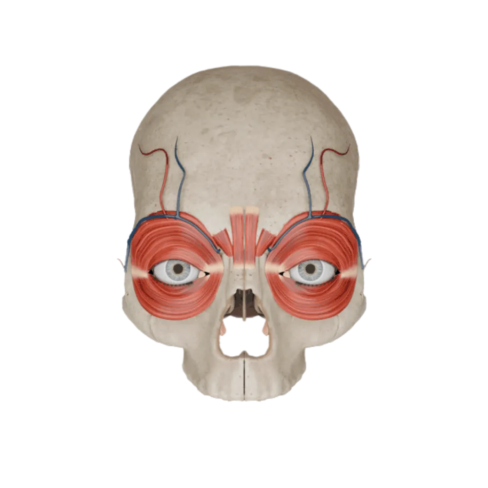
5. Which structure passes through the superior orbital fissure (fissura orbitalis superior)?
-
Maxillary nerve
Through the superior orbital fissure pass the III, IV, VI (abducens) cranial nerves, and the first branch of the V nerve (ophthalmic nerve), as well as the superior ophthalmic vein.
-
Abducens nerve
Through the superior orbital fissure pass the III, IV, VI (abducens) cranial nerves, and the first branch of the V nerve (ophthalmic nerve), as well as the superior ophthalmic vein.
-
Ophthalmic artery
Through the superior orbital fissure pass the III, IV, VI (abducens) cranial nerves, and the first branch of the V nerve (ophthalmic nerve), as well as the superior ophthalmic vein.
-
Infraorbital nerve
Through the superior orbital fissure pass the III, IV, VI (abducens) cranial nerves, and the first branch of the V nerve (ophthalmic nerve), as well as the superior ophthalmic vein.
-
I find it difficult to answer
Through the superior orbital fissure pass the III, IV, VI (abducens) cranial nerves, and the first branch of the V nerve (ophthalmic nerve), as well as the superior ophthalmic vein.
6. Which structure exits the cranial cavity through the oval foramen (foramen ovale)?
-
Maxillary nerve
Through the foramen ovale, from the middle cranial fossa to the infratemporal fossa, the mandibular nerve (third branch of the trigeminal nerve) exits.
-
Facial nerve
Through the foramen ovale, from the middle cranial fossa to the infratemporal fossa, the mandibular nerve (third branch of the trigeminal nerve) exits.
-
Mandibular nerve
Through the foramen ovale, from the middle cranial fossa to the infratemporal fossa, the mandibular nerve (third branch of the trigeminal nerve) exits.
-
Vagus nerve
Through the foramen ovale, from the middle cranial fossa to the infratemporal fossa, the mandibular nerve (third branch of the trigeminal nerve) exits.
-
I find it difficult to answer
Through the foramen ovale, from the middle cranial fossa to the infratemporal fossa, the mandibular nerve (third branch of the trigeminal nerve) exits.
7. Which nerves enter the internal auditory meatus (porus acusticus internus)?
-
Trigeminal and abducent
Into the internal auditory canal go the facial (VII cranial nerve) and vestibulocochlear (VIII cranial nerve) nerves.
-
Glossopharyngeal and vagus
Into the internal auditory canal go the facial (VII cranial nerve) and vestibulocochlear (VIII cranial nerve) nerves.
-
Facial and vestibulocochlear
Into the internal auditory canal go the facial (VII cranial nerve) and vestibulocochlear (VIII cranial nerve) nerves.
-
Trochlear and oculomotor
Into the internal auditory canal go the facial (VII cranial nerve) and vestibulocochlear (VIII cranial nerve) nerves.
-
I find it difficult to answer
Into the internal auditory canal go the facial (VII cranial nerve) and vestibulocochlear (VIII cranial nerve) nerves.
8. Which foramen marks the exit of the facial nerve (nervus facialis) from the temporal bone at the cranial base?
-
Foramen lacerum
The facial nerve exits the facial canal of the temporal bone through the stylomastoid foramen
-
Stylomastoid foramen
The facial nerve exits the facial canal of the temporal bone through the stylomastoid foramen
-
Jugular foramen
The facial nerve exits the facial canal of the temporal bone through the stylomastoid foramen
-
Mastoid foramen
The facial nerve exits the facial canal of the temporal bone through the stylomastoid foramen
-
I find it difficult to answer
The facial nerve exits the facial canal of the temporal bone through the stylomastoid foramen
9. Where is the external opening of the carotid canal (apertura externa canalis carotici) through which the internal carotid artery enters located?
-
On the inferior surface of the temporal bone pyramid
The external opening of the carotid canal is located on the inferior surface of the petrous part of the temporal bone
-
In the region of the greater wing of the sphenoid bone
The external opening of the carotid canal is located on the inferior surface of the petrous part of the temporal bone
-
On the basilar part of the occipital bone
The external opening of the carotid canal is located on the inferior surface of the petrous part of the temporal bone
-
On the anterior surface of the petrous part of the temporal bone
The external opening of the carotid canal is located on the inferior surface of the petrous part of the temporal bone
-
I find it difficult to answer
The external opening of the carotid canal is located on the inferior surface of the petrous part of the temporal bone
10. Which of the listed structures does NOT pass through the foramen magnum?
-
Vertebral artery
The internal jugular vein forms at the jugular foramen, and through the foramen magnum pass the medulla oblongata, vertebral arteries, and roots of the XI nerve.
-
Spinal roots of the accessory nerve
The internal jugular vein forms at the jugular foramen, and through the foramen magnum pass the medulla oblongata, vertebral arteries, and roots of the XI nerve.
-
Internal jugular vein
The internal jugular vein forms at the jugular foramen, and through the foramen magnum pass the medulla oblongata, vertebral arteries, and roots of the XI nerve.
-
Medulla oblongata
The internal jugular vein forms at the jugular foramen, and through the foramen magnum pass the medulla oblongata, vertebral arteries, and roots of the XI nerve.
-
I find it difficult to answer
The internal jugular vein forms at the jugular foramen, and through the foramen magnum pass the medulla oblongata, vertebral arteries, and roots of the XI nerve.
11. Which foramen of the cranial base transmits the olfactory nerves (nervi olfactorii)?
-
Crista galli
The olfactory nerves (I cranial nerves) enter the anterior cranial fossa through the foramina of the lamina cribrosa of the ethmoid bone.
-
Perpendicular plate of the ethmoid bone
The olfactory nerves (I cranial nerves) enter the anterior cranial fossa through the foramina of the lamina cribrosa of the ethmoid bone.
-
Smooth part of frontal bone
The olfactory nerves (I cranial nerves) enter the anterior cranial fossa through the foramina of the lamina cribrosa of the ethmoid bone.
-
Cribriform plate of the ethmoid bone
The olfactory nerves (I cranial nerves) enter the anterior cranial fossa through the foramina of the lamina cribrosa of the ethmoid bone.
-
I find it difficult to answer
The olfactory nerves (I cranial nerves) enter the anterior cranial fossa through the foramina of the lamina cribrosa of the ethmoid bone.
12. Which structures pass through the pterygoid (vidian) canal of the sphenoid bone?
-
Greater and lesser palatine nerves
In the pterygoid canal (canalis pterygoideus), the nerve of the pterygoid canal (formed by the union of the greater and deep petrosal nerves) and vessels pass through.
-
Maxillary artery and vein
In the pterygoid canal (canalis pterygoideus), the nerve of the pterygoid canal (formed by the union of the greater and deep petrosal nerves) and vessels pass through.
-
Homonymous nerve and vessels
In the pterygoid canal (canalis pterygoideus), the nerve of the pterygoid canal (formed by the union of the greater and deep petrosal nerves) and vessels pass through.
-
Pterygopalatine ganglion
In the pterygoid canal (canalis pterygoideus), the nerve of the pterygoid canal (formed by the union of the greater and deep petrosal nerves) and vessels pass through.
-
I find it difficult to answer
In the pterygoid canal (canalis pterygoideus), the nerve of the pterygoid canal (formed by the union of the greater and deep petrosal nerves) and vessels pass through.
13. What is the functional purpose of the condylar canal (canalis condylaris)?
-
Passage of the accessory nerve
The condylar canal is an inconstant structure containing the condylar emissary vein, connecting the sigmoid sinus with external veins.
-
Passage of the emissary vein
The condylar canal is an inconstant structure containing the condylar emissary vein, connecting the sigmoid sinus with external veins.
-
Passage of the occipital artery
The condylar canal is an inconstant structure containing the condylar emissary vein, connecting the sigmoid sinus with external veins.
-
Exit of the hypoglossal nerve
The condylar canal is an inconstant structure containing the condylar emissary vein, connecting the sigmoid sinus with external veins.
-
I find it difficult to answer
The condylar canal is an inconstant structure containing the condylar emissary vein, connecting the sigmoid sinus with external veins.
14. Which structure is adjacent to the internal aperture of the carotid canal in the region of the foramen lacerum in a living person?
-
Dura mater without cartilaginous tissue
In a living person, the foramen lacerum is closed by synchondrosis (fibrocartilage), on the upper surface of which the internal carotid artery runs.
-
Mandibular nerve
In a living person, the foramen lacerum is closed by synchondrosis (fibrocartilage), on the upper surface of which the internal carotid artery runs.
-
Glossopharyngeal nerve
In a living person, the foramen lacerum is closed by synchondrosis (fibrocartilage), on the upper surface of which the internal carotid artery runs.
-
Fibrocartilage
In a living person, the foramen lacerum is closed by synchondrosis (fibrocartilage), on the upper surface of which the internal carotid artery runs.
-
I find it difficult to answer
In a living person, the foramen lacerum is closed by synchondrosis (fibrocartilage), on the upper surface of which the internal carotid artery runs.
15. Where is the hypoglossal canal (canalis hypoglossi) located?
-
In the lesser wings of the sphenoid bone
The hypoglossal canal pierces the base of the occipital condyle and serves as the exit for the XII cranial nerve pair.
-
At the apex of the temporal bone pyramid
The hypoglossal canal pierces the base of the occipital condyle and serves as the exit for the XII cranial nerve pair.
-
In the body of the sphenoid bone
The hypoglossal canal pierces the base of the occipital condyle and serves as the exit for the XII cranial nerve pair.
-
At the base of the occipital condyle
The hypoglossal canal pierces the base of the occipital condyle and serves as the exit for the XII cranial nerve pair.
-
I find it difficult to answer
The hypoglossal canal pierces the base of the occipital condyle and serves as the exit for the XII cranial nerve pair.
16. What communication does the inferior orbital fissure (fissura orbitalis inferior) provide?
-
Orbits with the anterior cranial fossa
The inferior orbital fissure connects the orbital cavity with the pterygopalatine and infratemporal fossae, transmitting the infraorbital and zygomatic nerves.
-
Orbits with the pterygopalatine and infratemporal fossae
The inferior orbital fissure connects the orbital cavity with the pterygopalatine and infratemporal fossae, transmitting the infraorbital and zygomatic nerves.
-
Orbits with the middle cranial fossa
The inferior orbital fissure connects the orbital cavity with the pterygopalatine and infratemporal fossae, transmitting the infraorbital and zygomatic nerves.
-
Orbits with the nasal cavity
The inferior orbital fissure connects the orbital cavity with the pterygopalatine and infratemporal fossae, transmitting the infraorbital and zygomatic nerves.
-
I find it difficult to answer
The inferior orbital fissure connects the orbital cavity with the pterygopalatine and infratemporal fossae, transmitting the infraorbital and zygomatic nerves.
17. Which blood vessel passes together with cranial nerve pairs VII and VIII through the internal auditory canal?
-
Anterior inferior cerebellar artery
The labyrinthine artery (a branch of the basilar artery) enters the internal auditory canal along with the facial and vestibulocochlear nerves.
-
Posterior cerebral artery
The labyrinthine artery (a branch of the basilar artery) enters the internal auditory canal along with the facial and vestibulocochlear nerves.
-
Labyrinthine artery
The labyrinthine artery (a branch of the basilar artery) enters the internal auditory canal along with the facial and vestibulocochlear nerves.
-
Middle meningeal artery
The labyrinthine artery (a branch of the basilar artery) enters the internal auditory canal along with the facial and vestibulocochlear nerves.
-
I find it difficult to answer
The labyrinthine artery (a branch of the basilar artery) enters the internal auditory canal along with the facial and vestibulocochlear nerves.
18. Where does the greater palatine foramen (foramen palatinum majus) on the hard palate open?
-
Into the pterygopalatine fossa through the greater palatine canal
The greater palatine foramen serves as the exit of the greater palatine canal, which connects the oral cavity with the pterygopalatine fossa.
-
Into the nasal cavity
The greater palatine foramen serves as the exit of the greater palatine canal, which connects the oral cavity with the pterygopalatine fossa.
-
Into the infratemporal fossa
The greater palatine foramen serves as the exit of the greater palatine canal, which connects the oral cavity with the pterygopalatine fossa.
-
Into the maxillary sinus
The greater palatine foramen serves as the exit of the greater palatine canal, which connects the oral cavity with the pterygopalatine fossa.
-
I find it difficult to answer
The greater palatine foramen serves as the exit of the greater palatine canal, which connects the oral cavity with the pterygopalatine fossa.
19. What exits through the hiatus of the canal for the greater petrosal nerve (hiatus canalis nervi petrosi majoris)?
-
Lesser petrosal nerve
Through this hiatus on the anterior surface of the temporal bone pyramid exits the greater petrosal nerve (a branch of the facial nerve).
-
Chorda tympani
Through this hiatus on the anterior surface of the temporal bone pyramid exits the greater petrosal nerve (a branch of the facial nerve).
-
Auriculotemporal nerve
Through this hiatus on the anterior surface of the temporal bone pyramid exits the greater petrosal nerve (a branch of the facial nerve).
-
Greater petrosal nerve
Through this hiatus on the anterior surface of the temporal bone pyramid exits the greater petrosal nerve (a branch of the facial nerve).
-
I find it difficult to answer
Through this hiatus on the anterior surface of the temporal bone pyramid exits the greater petrosal nerve (a branch of the facial nerve).
20. Which structure is located in the foramen cecum of the frontal bone?
-
Emissary vein connecting the nasal vein with the superior sagittal sinus
The foramen cecum often contains an emissary vein that provides venous drainage from the nasal mucosa to the superior sagittal sinus.
-
Olfactory bulb
The foramen cecum often contains an emissary vein that provides venous drainage from the nasal mucosa to the superior sagittal sinus.
-
Optic chiasm
The foramen cecum often contains an emissary vein that provides venous drainage from the nasal mucosa to the superior sagittal sinus.
-
Ophthalmic artery
The foramen cecum often contains an emissary vein that provides venous drainage from the nasal mucosa to the superior sagittal sinus.
-
I find it difficult to answer
The foramen cecum often contains an emissary vein that provides venous drainage from the nasal mucosa to the superior sagittal sinus.
Retake this quiz?
Your current progress will be reset.