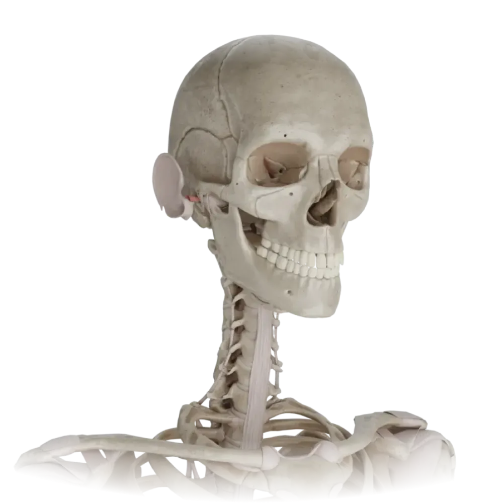
Test on the anatomy of the bones of the head and neck
Assess knowledge of head and neck bones. The test assesses the topography of the orbit, fossae, cranial canals, nasal cavity, and cervical vertebrae.
1/20
0%
bold
text
1. Which foramen connects the pterygopalatine fossa with the nasal cavity?
Foramen rotundum
The pterygopalatine fossa communicates with the nasal cavity through the sphenopalatine foramen (foramen sphenopalatinum).
Sphenopalatine foramen
The pterygopalatine fossa communicates with the nasal cavity through the sphenopalatine foramen (foramen sphenopalatinum).
Inferior orbital fissure
The pterygopalatine fossa communicates with the nasal cavity through the sphenopalatine foramen (foramen sphenopalatinum).
Pterygoid canal
The pterygopalatine fossa communicates with the nasal cavity through the sphenopalatine foramen (foramen sphenopalatinum).
I find it difficult to answer
The pterygopalatine fossa communicates with the nasal cavity through the sphenopalatine foramen (foramen sphenopalatinum).
2. Which structure does NOT pass through the jugular foramen of the skull?
Vagus nerve
The hypoglossal nerve (cranial nerve XII) passes through its own canal, rather than the jugular foramen.
Accessory nerve
The hypoglossal nerve (cranial nerve XII) passes through its own canal, rather than the jugular foramen.
Glossopharyngeal nerve
The hypoglossal nerve (cranial nerve XII) passes through its own canal, rather than the jugular foramen.
Hypoglossal nerve
The hypoglossal nerve (cranial nerve XII) passes through its own canal, rather than the jugular foramen.
I find it difficult to answer
The hypoglossal nerve (cranial nerve XII) passes through its own canal, rather than the jugular foramen.
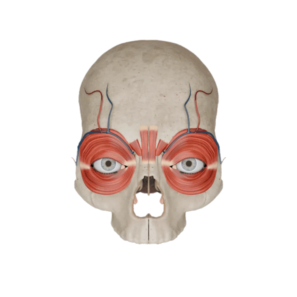
3. Which bones form the superior wall of the orbit?
Greater wing of sphenoid and zygomatic bone
The superior wall of the orbit is formed by the orbital part of the frontal bone and the lesser wing of the sphenoid bone.
Orbital plate of the ethmoid bone and lacrimal bone
The superior wall of the orbit is formed by the orbital part of the frontal bone and the lesser wing of the sphenoid bone.
Orbital part of frontal bone and lesser wing of sphenoid
The superior wall of the orbit is formed by the orbital part of the frontal bone and the lesser wing of the sphenoid bone.
Maxilla and palatine bone
The superior wall of the orbit is formed by the orbital part of the frontal bone and the lesser wing of the sphenoid bone.
I find it difficult to answer
The superior wall of the orbit is formed by the orbital part of the frontal bone and the lesser wing of the sphenoid bone.
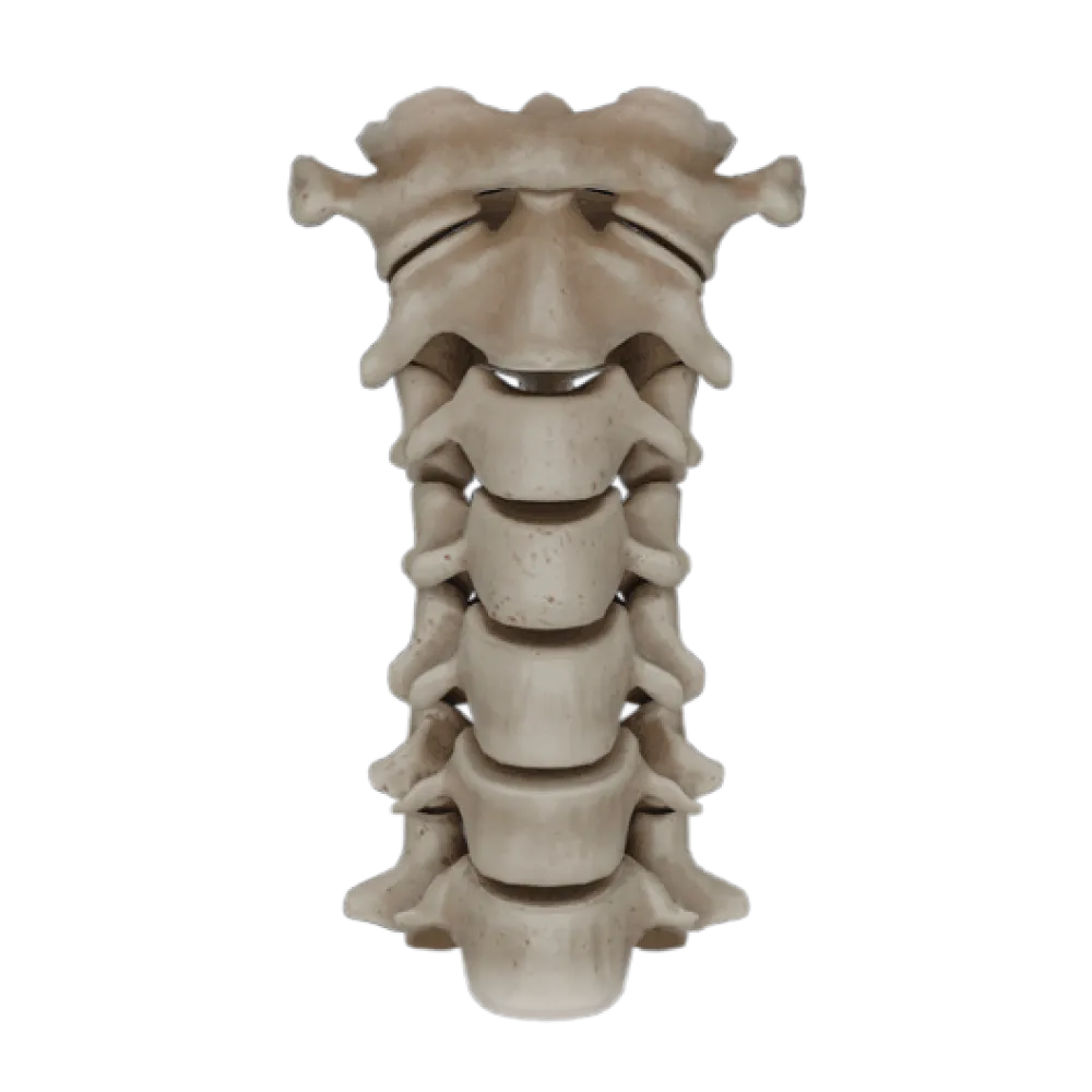
4. What anatomical feature is a specific distinguishing sign of the second cervical vertebra (axis)?
Bifid spinous process
The dens is a specific feature of the axis, acting as the pivot for rotation of the atlas.
Transverse process foramina
The dens is a specific feature of the axis, acting as the pivot for rotation of the atlas.
Dens
The dens is a specific feature of the axis, acting as the pivot for rotation of the atlas.
Anterior and posterior tubercles on transverse processes
The dens is a specific feature of the axis, acting as the pivot for rotation of the atlas.
I find it difficult to answer
The dens is a specific feature of the axis, acting as the pivot for rotation of the atlas.
5. Into which anatomical area does the foramen rotundum lead from the middle cranial fossa?
Pterygopalatine fossa
The foramen rotundum opens into the pterygopalatine fossa, allowing the passage of the maxillary nerve.
Orbit
The foramen rotundum opens into the pterygopalatine fossa, allowing the passage of the maxillary nerve.
Nasal cavity
The foramen rotundum opens into the pterygopalatine fossa, allowing the passage of the maxillary nerve.
Infratemporal fossa
The foramen rotundum opens into the pterygopalatine fossa, allowing the passage of the maxillary nerve.
I find it difficult to answer
The foramen rotundum opens into the pterygopalatine fossa, allowing the passage of the maxillary nerve.
6. Into which nasal meatus does the maxillary sinus open?
Superior nasal meatus
The maxillary sinus opens through the semilunar hiatus into the middle nasal meatus.
Middle nasal meatus
The maxillary sinus opens through the semilunar hiatus into the middle nasal meatus.
Inferior nasal meatus
The maxillary sinus opens through the semilunar hiatus into the middle nasal meatus.
Common nasal meatus
The maxillary sinus opens through the semilunar hiatus into the middle nasal meatus.
I find it difficult to answer
The maxillary sinus opens through the semilunar hiatus into the middle nasal meatus.
7. Which bony structure forms the medial wall of the infratemporal fossa?
Lateral plate of the pterygoid process of the sphenoid bone
The medial wall of the infratemporal fossa is formed by the lateral plate of the pterygoid process of the sphenoid bone.
Maxillary tuberosity
The medial wall of the infratemporal fossa is formed by the lateral plate of the pterygoid process of the sphenoid bone.
Mandibular ramus
The medial wall of the infratemporal fossa is formed by the lateral plate of the pterygoid process of the sphenoid bone.
Greater wing of sphenoid bone
The medial wall of the infratemporal fossa is formed by the lateral plate of the pterygoid process of the sphenoid bone.
I find it difficult to answer
The medial wall of the infratemporal fossa is formed by the lateral plate of the pterygoid process of the sphenoid bone.
8. Which cranial nerves pass through the internal acoustic meatus?
III, IV, VI pairs
Through the internal acoustic meatus follow the facial (VII) and vestibulocochlear (VIII) nerves.
VII and VIII pairs
Through the internal acoustic meatus follow the facial (VII) and vestibulocochlear (VIII) nerves.
IX, X, XI pairs
Through the internal acoustic meatus follow the facial (VII) and vestibulocochlear (VIII) nerves.
V and VII pairs
Through the internal acoustic meatus follow the facial (VII) and vestibulocochlear (VIII) nerves.
I find it difficult to answer
Through the internal acoustic meatus follow the facial (VII) and vestibulocochlear (VIII) nerves.
9. Through which formation does the orbit communicate with the middle cranial fossa?
Inferior orbital fissure and foramen rotundum
The orbit communicates with the middle cranial fossa via the optic canal and the superior orbital fissure.
Nasolacrimal canal and anterior ethmoidal foramen
The orbit communicates with the middle cranial fossa via the optic canal and the superior orbital fissure.
Sphenopalatine foramen and optic canal
The orbit communicates with the middle cranial fossa via the optic canal and the superior orbital fissure.
Optic canal and superior orbital fissure
The orbit communicates with the middle cranial fossa via the optic canal and the superior orbital fissure.
I find it difficult to answer
The orbit communicates with the middle cranial fossa via the optic canal and the superior orbital fissure.
10. Which foramen serves as the entrance for the carotid canal of the temporal bone?
Internal carotid foramen at the apex of the petrous part
The carotid canal begins at the external carotid foramen (foramen caroticum externum) on the inferior surface of the petrous part of the temporal bone.
Foramen spinosum on the greater wing of the sphenoid bone
The carotid canal begins at the external carotid foramen (foramen caroticum externum) on the inferior surface of the petrous part of the temporal bone.
External carotid foramen on the inferior surface of the petrous part
The carotid canal begins at the external carotid foramen (foramen caroticum externum) on the inferior surface of the petrous part of the temporal bone.
Foramen lacerum
The carotid canal begins at the external carotid foramen (foramen caroticum externum) on the inferior surface of the petrous part of the temporal bone.
I find it difficult to answer
The carotid canal begins at the external carotid foramen (foramen caroticum externum) on the inferior surface of the petrous part of the temporal bone.
11. Which structure is absent in the first cervical vertebra (atlas)?
Posterior arch
The atlas (C1) lacks a vertebral body, which fuses to the axis during embryogenic development to form the dens.
Lateral masses
The atlas (C1) lacks a vertebral body, which fuses to the axis during embryogenic development to form the dens.
Transverse process foramina
The atlas (C1) lacks a vertebral body, which fuses to the axis during embryogenic development to form the dens.
Vertebral body
The atlas (C1) lacks a vertebral body, which fuses to the axis during embryogenic development to form the dens.
I find it difficult to answer
The atlas (C1) lacks a vertebral body, which fuses to the axis during embryogenic development to form the dens.
12. What passes through the oval foramen (foramen ovale) of the sphenoid bone?
Ophthalmic nerve
Through the foramen ovale exits the mandibular nerve (third branch of the trigeminal nerve) from the cranial cavity.
Middle meningeal artery
Through the foramen ovale exits the mandibular nerve (third branch of the trigeminal nerve) from the cranial cavity.
Maxillary nerve
Through the foramen ovale exits the mandibular nerve (third branch of the trigeminal nerve) from the cranial cavity.
Mandibular nerve
Through the foramen ovale exits the mandibular nerve (third branch of the trigeminal nerve) from the cranial cavity.
I find it difficult to answer
Through the foramen ovale exits the mandibular nerve (third branch of the trigeminal nerve) from the cranial cavity.
13. Which ganglion is located in the pterygopalatine fossa?
Otic ganglion
In the pterygopalatine fossa is located the parasympathetic pterygopalatine ganglion (ganglion pterygopalatinum).
Pterygopalatine ganglion
In the pterygopalatine fossa is located the parasympathetic pterygopalatine ganglion (ganglion pterygopalatinum).
Ciliary ganglion
In the pterygopalatine fossa is located the parasympathetic pterygopalatine ganglion (ganglion pterygopalatinum).
Submandibular ganglion
In the pterygopalatine fossa is located the parasympathetic pterygopalatine ganglion (ganglion pterygopalatinum).
I find it difficult to answer
In the pterygopalatine fossa is located the parasympathetic pterygopalatine ganglion (ganglion pterygopalatinum).
14. In which part of the nasal cavity does the nasolacrimal canal open?
Superior nasal meatus
The nasolacrimal canal (canalis nasolacrimalis) opens in the anterior part of the inferior nasal meatus.
Middle nasal meatus
The nasolacrimal canal (canalis nasolacrimalis) opens in the anterior part of the inferior nasal meatus.
Inferior nasal meatus
The nasolacrimal canal (canalis nasolacrimalis) opens in the anterior part of the inferior nasal meatus.
Sphenethmoidal recess
The nasolacrimal canal (canalis nasolacrimalis) opens in the anterior part of the inferior nasal meatus.
I find it difficult to answer
The nasolacrimal canal (canalis nasolacrimalis) opens in the anterior part of the inferior nasal meatus.
15. With which nasal meatus does the sphenoidal sinus communicate?
Superior nasal meatus (sphenoethmoidal recess)
The sphenoidal sinus opens by its aperture into the sphenoethmoidal recess of the superior nasal meatus.
Middle nasal meatus
The sphenoidal sinus opens by its aperture into the sphenoethmoidal recess of the superior nasal meatus.
Inferior nasal meatus
The sphenoidal sinus opens by its aperture into the sphenoethmoidal recess of the superior nasal meatus.
Common nasal meatus
The sphenoidal sinus opens by its aperture into the sphenoethmoidal recess of the superior nasal meatus.
I find it difficult to answer
The sphenoidal sinus opens by its aperture into the sphenoethmoidal recess of the superior nasal meatus.
16. Which bones contribute to the formation of the jugular foramen (foramen jugulare)?
Temporal and sphenoid bones
The jugular foramen is formed by the jugular notches of the occipital and temporal (petrous part) bones.
Occipital and parietal bones
The jugular foramen is formed by the jugular notches of the occipital and temporal (petrous part) bones.
Sphenoid and frontal bones
The jugular foramen is formed by the jugular notches of the occipital and temporal (petrous part) bones.
Occipital and temporal bones
The jugular foramen is formed by the jugular notches of the occipital and temporal (petrous part) bones.
I find it difficult to answer
The jugular foramen is formed by the jugular notches of the occipital and temporal (petrous part) bones.
17. Through which foramina of the cervical vertebrae does the vertebral artery normally pass?
Transverse process foramina from C6 to C1
The vertebral artery passes through the foramina transversaria of the cervical vertebrae, typically starting from the sixth (C6) to the atlas (C1).
Intervertebral foramina
The vertebral artery passes through the foramina transversaria of the cervical vertebrae, typically starting from the sixth (C6) to the atlas (C1).
Vertebral canal
The vertebral artery passes through the foramina transversaria of the cervical vertebrae, typically starting from the sixth (C6) to the atlas (C1).
Transverse process foramina from C7 to C1
The vertebral artery passes through the foramina transversaria of the cervical vertebrae, typically starting from the sixth (C6) to the atlas (C1).
I find it difficult to answer
The vertebral artery passes through the foramina transversaria of the cervical vertebrae, typically starting from the sixth (C6) to the atlas (C1).
18. With which fossae does the inferior orbital fissure connect the orbit?
With the temporal and infratemporal fossae
The inferior orbital fissure connects the orbit with the pterygopalatine fossa (in its posterior part) and with the infratemporal fossa (in its anterior part).
With the middle and anterior cranial fossae
The inferior orbital fissure connects the orbit with the pterygopalatine fossa (in its posterior part) and with the infratemporal fossa (in its anterior part).
With the pterygopalatine and infratemporal fossae
The inferior orbital fissure connects the orbit with the pterygopalatine fossa (in its posterior part) and with the infratemporal fossa (in its anterior part).
With the canine and pterygopalatine fossae
The inferior orbital fissure connects the orbit with the pterygopalatine fossa (in its posterior part) and with the infratemporal fossa (in its anterior part).
I find it difficult to answer
The inferior orbital fissure connects the orbit with the pterygopalatine fossa (in its posterior part) and with the infratemporal fossa (in its anterior part).
19. Which artery enters the cranial cavity through the foramen spinosum?
Anterior meningeal artery
Through the foramen spinosum of the greater wing of the sphenoid bone penetrates the middle meningeal artery into the middle cranial fossa.
Internal carotid artery
Through the foramen spinosum of the greater wing of the sphenoid bone penetrates the middle meningeal artery into the middle cranial fossa.
Middle meningeal artery
Through the foramen spinosum of the greater wing of the sphenoid bone penetrates the middle meningeal artery into the middle cranial fossa.
Vertebral artery
Through the foramen spinosum of the greater wing of the sphenoid bone penetrates the middle meningeal artery into the middle cranial fossa.
I find it difficult to answer
Through the foramen spinosum of the greater wing of the sphenoid bone penetrates the middle meningeal artery into the middle cranial fossa.
20. What structures form the hard palate (palatum osseum)?
Alveolar processes of the maxillae and perpendicular plates of the palatine bones
The hard bony palate is formed by the palatine processes of the maxillae (anteriorly) and the horizontal plates of the palatine bones (posteriorly).
Palatine processes of the maxillae and horizontal plates of the palatine bones
The hard bony palate is formed by the palatine processes of the maxillae (anteriorly) and the horizontal plates of the palatine bones (posteriorly).
Palatine processes of the maxillae and pterygoid processes of the sphenoid bone
The hard bony palate is formed by the palatine processes of the maxillae (anteriorly) and the horizontal plates of the palatine bones (posteriorly).
Horizontal plates of the palatine bones and vomer
The hard bony palate is formed by the palatine processes of the maxillae (anteriorly) and the horizontal plates of the palatine bones (posteriorly).
I find it difficult to answer
The hard bony palate is formed by the palatine processes of the maxillae (anteriorly) and the horizontal plates of the palatine bones (posteriorly).