
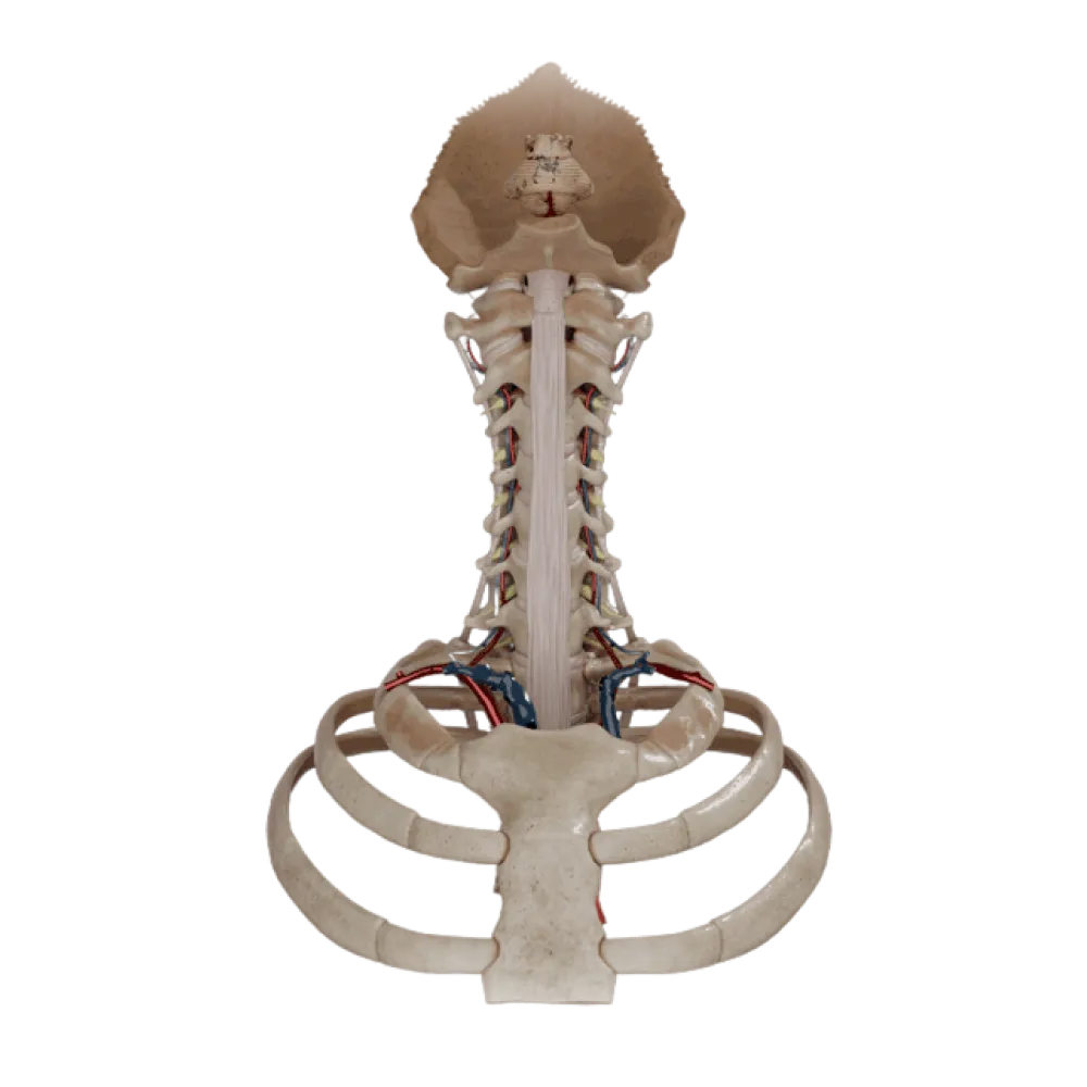
Vertebral artery anatomy test
Check your knowledge of vertebral artery anatomy. The test covers its topography, segments (V1-V4), branches, and areas of blood supply.
1/20
bold
text
1. Which artery does the vertebral artery arise from?
-
Common carotid artery
The vertebral artery is the first branch of the subclavian artery, arising from its superior semicircle.
-
Subclavian artery
The vertebral artery is the first branch of the subclavian artery, arising from its superior semicircle.
-
External carotid artery
The vertebral artery is the first branch of the subclavian artery, arising from its superior semicircle.
-
Thyrocervical trunk
The vertebral artery is the first branch of the subclavian artery, arising from its superior semicircle.
-
I find it difficult to answer
The vertebral artery is the first branch of the subclavian artery, arising from its superior semicircle.
2. Through the transverse foramen of which vertebra does the vertebral artery normally NOT pass?
-
Atlas (C1)
The vertebral artery typically enters the transverse foramen of C6, bypassing the transverse foramen of C7.
-
Axis (C2)
The vertebral artery typically enters the transverse foramen of C6, bypassing the transverse foramen of C7.
-
Sixth cervical vertebra (C6)
The vertebral artery typically enters the transverse foramen of C6, bypassing the transverse foramen of C7.
-
Seventh cervical vertebra (C7)
The vertebral artery typically enters the transverse foramen of C6, bypassing the transverse foramen of C7.
-
I find it difficult to answer
The vertebral artery typically enters the transverse foramen of C6, bypassing the transverse foramen of C7.
3. What is the name of the part of the vertebral artery (V3) located on the arch of the atlas?
-
Atlantic part
The atlantic part (V3) encircles the lateral mass of the atlas and lies in the corresponding groove on its posterior arch.
-
Cervical part
The atlantic part (V3) encircles the lateral mass of the atlas and lies in the corresponding groove on its posterior arch.
-
Prevertebral part
The atlantic part (V3) encircles the lateral mass of the atlas and lies in the corresponding groove on its posterior arch.
-
Intracranial part
The atlantic part (V3) encircles the lateral mass of the atlas and lies in the corresponding groove on its posterior arch.
-
I find it difficult to answer
The atlantic part (V3) encircles the lateral mass of the atlas and lies in the corresponding groove on its posterior arch.
4. Which membrane does the vertebral artery pierce while entering the vertebral canal?
-
Tectorial membrane
After passing the groove of the atlas, the artery pierces the posterior atlanto-occipital membrane and the dura mater.
-
Anterior atlanto-occipital membrane
After passing the groove of the atlas, the artery pierces the posterior atlanto-occipital membrane and the dura mater.
-
Posterior atlanto-occipital membrane
After passing the groove of the atlas, the artery pierces the posterior atlanto-occipital membrane and the dura mater.
-
Nuchal ligament
After passing the groove of the atlas, the artery pierces the posterior atlanto-occipital membrane and the dura mater.
-
I find it difficult to answer
After passing the groove of the atlas, the artery pierces the posterior atlanto-occipital membrane and the dura mater.
5. Which segment of the vertebral artery (by classification) extends from its origin to the entry into the transverse foramen of C6?
-
V1 (Preforaminal part)
Segment V1 extends from the subclavian artery to the transverse foramen of the 6th cervical vertebra.
-
V2 (Cervical part)
Segment V1 extends from the subclavian artery to the transverse foramen of the 6th cervical vertebra.
-
V3 (Atlantic part)
Segment V1 extends from the subclavian artery to the transverse foramen of the 6th cervical vertebra.
-
V4 (Intracranial part)
Segment V1 extends from the subclavian artery to the transverse foramen of the 6th cervical vertebra.
-
I find it difficult to answer
Segment V1 extends from the subclavian artery to the transverse foramen of the 6th cervical vertebra.
6. Through which foramen does the vertebral artery enter the cranial cavity?
-
Jugular foramen
The intracranial part (V4) of the vertebral artery enters the cranial cavity through the foramen magnum.
-
Foramen magnum
The intracranial part (V4) of the vertebral artery enters the cranial cavity through the foramen magnum.
-
Foramen ovale
The intracranial part (V4) of the vertebral artery enters the cranial cavity through the foramen magnum.
-
Foramen lacerum
The intracranial part (V4) of the vertebral artery enters the cranial cavity through the foramen magnum.
-
I find it difficult to answer
The intracranial part (V4) of the vertebral artery enters the cranial cavity through the foramen magnum.
7. Which branch originates from the intracranial part of the vertebral artery?
-
Superior cerebellar artery
The posterior inferior cerebellar artery (PICA) is the largest branch of the intracranial part of the vertebral artery.
-
Anterior inferior cerebellar artery
The posterior inferior cerebellar artery (PICA) is the largest branch of the intracranial part of the vertebral artery.
-
Labyrinthine artery
The posterior inferior cerebellar artery (PICA) is the largest branch of the intracranial part of the vertebral artery.
-
Posterior inferior cerebellar artery
The posterior inferior cerebellar artery (PICA) is the largest branch of the intracranial part of the vertebral artery.
-
I find it difficult to answer
The posterior inferior cerebellar artery (PICA) is the largest branch of the intracranial part of the vertebral artery.
8. Where do the right and left vertebral arteries converge to form the basilar artery?
-
At the level of the corpus callosum
The vertebral arteries fuse into the basilar artery at the level of the posterior (inferior) border of the pons.
-
At the anterior border of the pons
The vertebral arteries fuse into the basilar artery at the level of the posterior (inferior) border of the pons.
-
At the posterior (inferior) border of the pons
The vertebral arteries fuse into the basilar artery at the level of the posterior (inferior) border of the pons.
-
In the interpeduncular fossa
The vertebral arteries fuse into the basilar artery at the level of the posterior (inferior) border of the pons.
-
I find it difficult to answer
The vertebral arteries fuse into the basilar artery at the level of the posterior (inferior) border of the pons.
9. Which artery is formed by the fusion of two branches from both vertebral arteries and descends along the anterior median fissure of the spinal cord?
-
Anterior spinal artery
The anterior spinal artery is formed by the fusion of two roots originating from the intracranial parts of the vertebral arteries.
-
Posterior spinal artery
The anterior spinal artery is formed by the fusion of two roots originating from the intracranial parts of the vertebral arteries.
-
Great radicular artery
The anterior spinal artery is formed by the fusion of two roots originating from the intracranial parts of the vertebral arteries.
-
Basilar artery
The anterior spinal artery is formed by the fusion of two roots originating from the intracranial parts of the vertebral arteries.
-
I find it difficult to answer
The anterior spinal artery is formed by the fusion of two roots originating from the intracranial parts of the vertebral arteries.
10. Between which muscles does the V1 segment of the vertebral artery pass?
-
Between the anterior and middle scalene muscles
The prevertebral part of the artery passes behind the common carotid artery between the anterior scalene muscle and the longus colli muscle.
-
Between the anterior scalene and longus colli muscles
The prevertebral part of the artery passes behind the common carotid artery between the anterior scalene muscle and the longus colli muscle.
-
Between the longus capitis and longus colli muscles
The prevertebral part of the artery passes behind the common carotid artery between the anterior scalene muscle and the longus colli muscle.
-
Between the middle and posterior scalene muscles
The prevertebral part of the artery passes behind the common carotid artery between the anterior scalene muscle and the longus colli muscle.
-
I find it difficult to answer
The prevertebral part of the artery passes behind the common carotid artery between the anterior scalene muscle and the longus colli muscle.
11. What accompanies the vertebral artery in the transverse foramina of the cervical vertebrae?
-
Vagus nerve
Around the artery in the vertebral artery canal, a venous plexus forms, and the vertebral nerve (sympathetic plexus) is located.
-
Internal jugular vein
Around the artery in the vertebral artery canal, a venous plexus forms, and the vertebral nerve (sympathetic plexus) is located.
-
Sympathetic trunk
Around the artery in the vertebral artery canal, a venous plexus forms, and the vertebral nerve (sympathetic plexus) is located.
-
Vertebral vein and sympathetic plexus
Around the artery in the vertebral artery canal, a venous plexus forms, and the vertebral nerve (sympathetic plexus) is located.
-
I find it difficult to answer
Around the artery in the vertebral artery canal, a venous plexus forms, and the vertebral nerve (sympathetic plexus) is located.
12. Which branches originate from the cervical part (V2) of the vertebral artery?
-
Meningeal branches
From segment V2, spinal branches (through intervertebral foramina) and muscular branches to the deep muscles of the neck arise.
-
Cerebral branches
From segment V2, spinal branches (through intervertebral foramina) and muscular branches to the deep muscles of the neck arise.
-
Spinal and muscular branches
From segment V2, spinal branches (through intervertebral foramina) and muscular branches to the deep muscles of the neck arise.
-
Bulbar branches
From segment V2, spinal branches (through intervertebral foramina) and muscular branches to the deep muscles of the neck arise.
-
I find it difficult to answer
From segment V2, spinal branches (through intervertebral foramina) and muscular branches to the deep muscles of the neck arise.
13. From where do posterior spinal arteries usually originate?
-
From the intracranial part of the vertebral artery or the posterior inferior cerebellar artery
Posterior spinal arteries are most commonly branches of the vertebral artery (V4) or the posterior inferior cerebellar artery.
-
From the basilar artery
Posterior spinal arteries are most commonly branches of the vertebral artery (V4) or the posterior inferior cerebellar artery.
-
From the anterior spinal artery
Posterior spinal arteries are most commonly branches of the vertebral artery (V4) or the posterior inferior cerebellar artery.
-
From the ascending cervical artery
Posterior spinal arteries are most commonly branches of the vertebral artery (V4) or the posterior inferior cerebellar artery.
-
I find it difficult to answer
Posterior spinal arteries are most commonly branches of the vertebral artery (V4) or the posterior inferior cerebellar artery.
14. Which structure is closely associated with the vertebral artery groove on the atlas?
-
Dens of the axis
The groove of the vertebral artery (sulcus arteriae vertebralis) is located on the superior surface of the posterior arch of the atlas.
-
Lateral mass of the atlas
The groove of the vertebral artery (sulcus arteriae vertebralis) is located on the superior surface of the posterior arch of the atlas.
-
Anterior arch of the atlas
The groove of the vertebral artery (sulcus arteriae vertebralis) is located on the superior surface of the posterior arch of the atlas.
-
Posterior arch of the atlas
The groove of the vertebral artery (sulcus arteriae vertebralis) is located on the superior surface of the posterior arch of the atlas.
-
I find it difficult to answer
The groove of the vertebral artery (sulcus arteriae vertebralis) is located on the superior surface of the posterior arch of the atlas.
15. Which area is supplied by the posterior inferior cerebellar artery?
-
Superior surface of the hemispheres of the cerebellum
The PICA supplies the posteroinferior surface of the cerebellum, the choroid plexus of the fourth ventricle, and the lateral portions of the medulla oblongata.
-
Posterior part of the inferior surface of the cerebellum and the medulla oblongata
The PICA supplies the posteroinferior surface of the cerebellum, the choroid plexus of the fourth ventricle, and the lateral portions of the medulla oblongata.
-
Anterior part of the inferior surface of the cerebellum
The PICA supplies the posteroinferior surface of the cerebellum, the choroid plexus of the fourth ventricle, and the lateral portions of the medulla oblongata.
-
Pons Varolii
The PICA supplies the posteroinferior surface of the cerebellum, the choroid plexus of the fourth ventricle, and the lateral portions of the medulla oblongata.
-
I find it difficult to answer
The PICA supplies the posteroinferior surface of the cerebellum, the choroid plexus of the fourth ventricle, and the lateral portions of the medulla oblongata.
16. How is the vertebral artery positioned in relation to the trunks of the brachial plexus?
-
Posterior to the trunks
In the neck, the vertebral artery is positioned anterior and medial to the trunks of the brachial plexus.
-
Pierces the trunks
In the neck, the vertebral artery is positioned anterior and medial to the trunks of the brachial plexus.
-
Anterior and medial to the trunks
In the neck, the vertebral artery is positioned anterior and medial to the trunks of the brachial plexus.
-
Lateral to the trunks
In the neck, the vertebral artery is positioned anterior and medial to the trunks of the brachial plexus.
-
I find it difficult to answer
In the neck, the vertebral artery is positioned anterior and medial to the trunks of the brachial plexus.
17. Which meningeal layer covers the vertebral artery in its intracranial segment?
-
Arachnoid mater
In the cranial cavity, the vertebral artery is located in the subarachnoid space, covered by the arachnoid mater.
-
Periosteum
In the cranial cavity, the vertebral artery is located in the subarachnoid space, covered by the arachnoid mater.
-
Ligamenta flava
In the cranial cavity, the vertebral artery is located in the subarachnoid space, covered by the arachnoid mater.
-
Tectorial membrane
In the cranial cavity, the vertebral artery is located in the subarachnoid space, covered by the arachnoid mater.
-
I find it difficult to answer
In the cranial cavity, the vertebral artery is located in the subarachnoid space, covered by the arachnoid mater.
18. In which neck triangle is the origin of the vertebral artery projected?
-
Submandibular triangle
The vertebral artery originates in the scalene-vertebral triangle, bounded by the longus colli and anterior scalene muscles.
-
Carotid triangle
The vertebral artery originates in the scalene-vertebral triangle, bounded by the longus colli and anterior scalene muscles.
-
Omotracheal triangle
The vertebral artery originates in the scalene-vertebral triangle, bounded by the longus colli and anterior scalene muscles.
-
Scalene-vertebral triangle
The vertebral artery originates in the scalene-vertebral triangle, bounded by the longus colli and anterior scalene muscles.
-
I find it difficult to answer
The vertebral artery originates in the scalene-vertebral triangle, bounded by the longus colli and anterior scalene muscles.
19. Which branches arise from the V4 segment of the vertebral artery to the medulla oblongata?
-
Pontine branches
The intracranial part of the vertebral artery gives off short bulbar branches to supply the medulla oblongata.
-
Bulbar (medullary) branches
The intracranial part of the vertebral artery gives off short bulbar branches to supply the medulla oblongata.
-
Thalamic branches
The intracranial part of the vertebral artery gives off short bulbar branches to supply the medulla oblongata.
-
Choroidal branches
The intracranial part of the vertebral artery gives off short bulbar branches to supply the medulla oblongata.
-
I find it difficult to answer
The intracranial part of the vertebral artery gives off short bulbar branches to supply the medulla oblongata.
20. Where is the meningeal branch of the vertebral artery located?
-
In the anterior cranial fossa
The meningeal branch of the vertebral artery originates in the area of the foramen magnum and supplies the dura mater of the posterior cranial fossa.
-
In the middle cranial fossa
The meningeal branch of the vertebral artery originates in the area of the foramen magnum and supplies the dura mater of the posterior cranial fossa.
-
In the posterior cranial fossa
The meningeal branch of the vertebral artery originates in the area of the foramen magnum and supplies the dura mater of the posterior cranial fossa.
-
In the spinal canal
The meningeal branch of the vertebral artery originates in the area of the foramen magnum and supplies the dura mater of the posterior cranial fossa.
-
I find it difficult to answer
The meningeal branch of the vertebral artery originates in the area of the foramen magnum and supplies the dura mater of the posterior cranial fossa.
Retake this quiz?
Your current progress will be reset.