
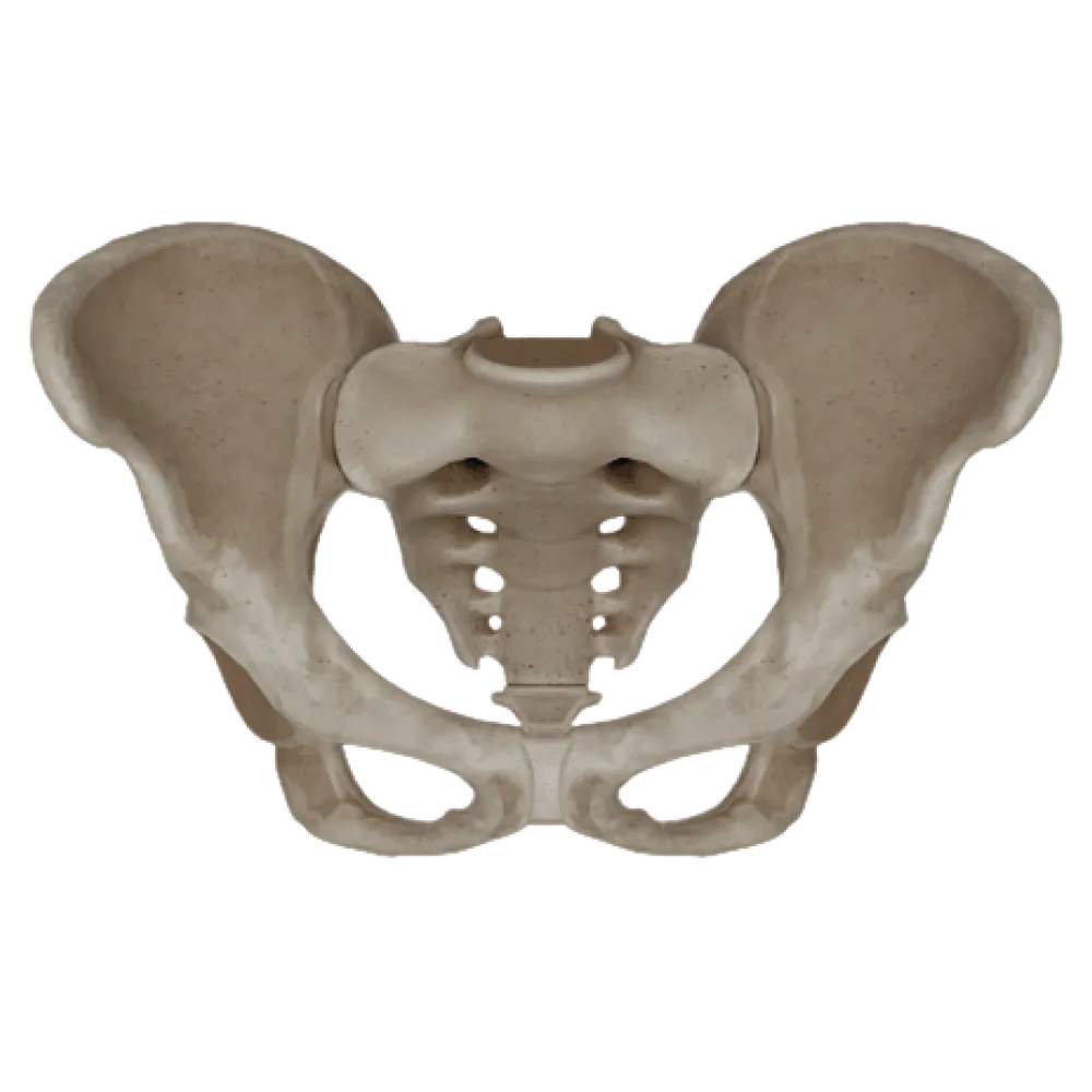
Test on the anatomy of the female bony pelvis
Evaluate your knowledge of the female bony pelvis anatomy. The test strictly examines the topography, dimensions, planes, and structural features in normal conditions.
1/20
bold
text
1. What is the normal size of the true conjugate (conjugata vera) of the female pelvis?
-
13 cm
The true (obstetric) conjugate is the distance from the sacral promontory to the most posterior point of the pubic symphysis, and it is normally about 11 cm.
-
10 cm
The true (obstetric) conjugate is the distance from the sacral promontory to the most posterior point of the pubic symphysis, and it is normally about 11 cm.
-
11 cm
The true (obstetric) conjugate is the distance from the sacral promontory to the most posterior point of the pubic symphysis, and it is normally about 11 cm.
-
12,5 cm
The true (obstetric) conjugate is the distance from the sacral promontory to the most posterior point of the pubic symphysis, and it is normally about 11 cm.
-
I find it difficult to answer
The true (obstetric) conjugate is the distance from the sacral promontory to the most posterior point of the pubic symphysis, and it is normally about 11 cm.
2. What is the normal distance between the anterior superior iliac spines (distantia spinarum)?
-
28-29 cm
Distantia spinarum — the distance between the anterior superior iliac spines, is normally 25-27 cm in women.
-
25-27 cm
Distantia spinarum — the distance between the anterior superior iliac spines, is normally 25-27 cm in women.
-
30-32 cm
Distantia spinarum — the distance between the anterior superior iliac spines, is normally 25-27 cm in women.
-
20-22 cm
Distantia spinarum — the distance between the anterior superior iliac spines, is normally 25-27 cm in women.
-
I find it difficult to answer
Distantia spinarum — the distance between the anterior superior iliac spines, is normally 25-27 cm in women.
3. What is the angle of the subpubic arch (angulus subpubicus) in a normal female pelvis?
-
70-75 degrees
Unlike the male pelvis (where the angle is acute, 70-75°), the female subpubic angle is obtuse and measures 90-100°.
-
50-60 degrees
Unlike the male pelvis (where the angle is acute, 70-75°), the female subpubic angle is obtuse and measures 90-100°.
-
90-100 degrees
Unlike the male pelvis (where the angle is acute, 70-75°), the female subpubic angle is obtuse and measures 90-100°.
-
110-120 degrees
Unlike the male pelvis (where the angle is acute, 70-75°), the female subpubic angle is obtuse and measures 90-100°.
-
I find it difficult to answer
Unlike the male pelvis (where the angle is acute, 70-75°), the female subpubic angle is obtuse and measures 90-100°.
4. The terminal line (linea terminalis) that separates the greater and lesser pelvis is formed by:
-
The wings of the sacrum, the inferior gluteal line, the pubic symphysis
The terminal line passes from the sacral promontory along the arcuate lines of the iliac bones, pubic crests, and the upper edge of the symphysis.
-
The promontory, arcuate line of the ilium, pubic crest, and upper edge of the symphysis
The terminal line passes from the sacral promontory along the arcuate lines of the iliac bones, pubic crests, and the upper edge of the symphysis.
-
The ischial spine and the sacrotuberous ligament
The terminal line passes from the sacral promontory along the arcuate lines of the iliac bones, pubic crests, and the upper edge of the symphysis.
-
The crest of the ilium and the anterior superior spine
The terminal line passes from the sacral promontory along the arcuate lines of the iliac bones, pubic crests, and the upper edge of the symphysis.
-
I find it difficult to answer
The terminal line passes from the sacral promontory along the arcuate lines of the iliac bones, pubic crests, and the upper edge of the symphysis.
5. The anatomical conjugate (conjugata anatomica) is approximately:
-
11,0 cm
The anatomical conjugate is measured from the promontory to the upper edge of the pubic symphysis and is normally about 11.5 cm.
-
12,5 cm
The anatomical conjugate is measured from the promontory to the upper edge of the pubic symphysis and is normally about 11.5 cm.
-
13,0 cm
The anatomical conjugate is measured from the promontory to the upper edge of the pubic symphysis and is normally about 11.5 cm.
-
11,5 cm
The anatomical conjugate is measured from the promontory to the upper edge of the pubic symphysis and is normally about 11.5 cm.
-
I find it difficult to answer
The anatomical conjugate is measured from the promontory to the upper edge of the pubic symphysis and is normally about 11.5 cm.
6. The diagonal conjugate (conjugata diagonalis) is the distance between:
-
The promontory and the lower edge of the pubic symphysis
The diagonal conjugate (normally about 13 cm) is measured during a vaginal examination from the lower edge of the symphysis to the sacral promontory.
-
The promontory and the upper edge of the pubic symphysis
The diagonal conjugate (normally about 13 cm) is measured during a vaginal examination from the lower edge of the symphysis to the sacral promontory.
-
The sacroiliac joint and the ilio-pubic eminence
The diagonal conjugate (normally about 13 cm) is measured during a vaginal examination from the lower edge of the symphysis to the sacral promontory.
-
The ischial spines
The diagonal conjugate (normally about 13 cm) is measured during a vaginal examination from the lower edge of the symphysis to the sacral promontory.
-
I find it difficult to answer
The diagonal conjugate (normally about 13 cm) is measured during a vaginal examination from the lower edge of the symphysis to the sacral promontory.
7. The transverse dimension of the true pelvis inlet (diameter transversa) is normally:
-
11 cm
The transverse dimension is the largest distance between the transverse lines of the opposite sides, measuring 13-13.5 cm.
-
12 cm
The transverse dimension is the largest distance between the transverse lines of the opposite sides, measuring 13-13.5 cm.
-
13 cm
The transverse dimension is the largest distance between the transverse lines of the opposite sides, measuring 13-13.5 cm.
-
14 cm
The transverse dimension is the largest distance between the transverse lines of the opposite sides, measuring 13-13.5 cm.
-
I find it difficult to answer
The transverse dimension is the largest distance between the transverse lines of the opposite sides, measuring 13-13.5 cm.
8. The external conjugate (conjugata externa) of the female pelvis is normally:
-
17-18 cm
The external conjugate is measured from the outer upper edge of the symphysis to the suprasacral fossa and normally measures 20-21 cm.
-
23-24 cm
The external conjugate is measured from the outer upper edge of the symphysis to the suprasacral fossa and normally measures 20-21 cm.
-
20-21 cm
The external conjugate is measured from the outer upper edge of the symphysis to the suprasacral fossa and normally measures 20-21 cm.
-
25-26 cm
The external conjugate is measured from the outer upper edge of the symphysis to the suprasacral fossa and normally measures 20-21 cm.
-
I find it difficult to answer
The external conjugate is measured from the outer upper edge of the symphysis to the suprasacral fossa and normally measures 20-21 cm.
9. The inclination angle of the pelvis (inclinatio pelvis) in women when standing is usually:
-
40-45°
The inclination angle of the pelvis (between the true pelvis inlet plane and the horizontal) in women is greater than in men, measuring 55-60°.
-
55-60°
The inclination angle of the pelvis (between the true pelvis inlet plane and the horizontal) in women is greater than in men, measuring 55-60°.
-
70-75°
The inclination angle of the pelvis (between the true pelvis inlet plane and the horizontal) in women is greater than in men, measuring 55-60°.
-
30-35°
The inclination angle of the pelvis (between the true pelvis inlet plane and the horizontal) in women is greater than in men, measuring 55-60°.
-
I find it difficult to answer
The inclination angle of the pelvis (between the true pelvis inlet plane and the horizontal) in women is greater than in men, measuring 55-60°.
10. The size of distantia cristarum in a normal female pelvis is:
-
25-27 cm
Distantia cristarum — the largest distance between the iliac crests, is normally 28-29 cm.
-
30-31 cm
Distantia cristarum — the largest distance between the iliac crests, is normally 28-29 cm.
-
28-29 cm
Distantia cristarum — the largest distance between the iliac crests, is normally 28-29 cm.
-
32-33 cm
Distantia cristarum — the largest distance between the iliac crests, is normally 28-29 cm.
-
I find it difficult to answer
Distantia cristarum — the largest distance between the iliac crests, is normally 28-29 cm.
11. The size of distantia trochanterica in a normal female pelvis is approximately:
-
28-29 cm
Distantia trochanterica — the distance between the greater trochanters of the femurs, is normally 30-32 cm.
-
33-35 cm
Distantia trochanterica — the distance between the greater trochanters of the femurs, is normally 30-32 cm.
-
25-27 cm
Distantia trochanterica — the distance between the greater trochanters of the femurs, is normally 30-32 cm.
-
30-32 cm
Distantia trochanterica — the distance between the greater trochanters of the femurs, is normally 30-32 cm.
-
I find it difficult to answer
Distantia trochanterica — the distance between the greater trochanters of the femurs, is normally 30-32 cm.
12. The anteroposterior dimension of the true pelvis outlet (diameter recta exitus pelvis) increases during childbirth due to:
-
The divergence of the pubic symphysis
Due to the mobility of the sacrococcygeal joint, the coccyx can deviate backward, increasing the anteroposterior dimension of the pelvic outlet by 1.5-2 cm.
-
The backward deviation of the coccyx
Due to the mobility of the sacrococcygeal joint, the coccyx can deviate backward, increasing the anteroposterior dimension of the pelvic outlet by 1.5-2 cm.
-
The rotations of the iliac bones
Due to the mobility of the sacrococcygeal joint, the coccyx can deviate backward, increasing the anteroposterior dimension of the pelvic outlet by 1.5-2 cm.
-
The flexion of the lumbar spine
Due to the mobility of the sacrococcygeal joint, the coccyx can deviate backward, increasing the anteroposterior dimension of the pelvic outlet by 1.5-2 cm.
-
I find it difficult to answer
Due to the mobility of the sacrococcygeal joint, the coccyx can deviate backward, increasing the anteroposterior dimension of the pelvic outlet by 1.5-2 cm.
13. The normal size of the anteroposterior dimension of the true pelvis outlet with a deviated coccyx is:
-
9,5 cm
In a typical state, the anteroposterior dimension is 9.5 cm, and with the backward deviation of the coccyx during childbirth, it increases to 11.5 cm.
-
10,5 cm
In a typical state, the anteroposterior dimension is 9.5 cm, and with the backward deviation of the coccyx during childbirth, it increases to 11.5 cm.
-
11,5 cm
In a typical state, the anteroposterior dimension is 9.5 cm, and with the backward deviation of the coccyx during childbirth, it increases to 11.5 cm.
-
12,5 cm
In a typical state, the anteroposterior dimension is 9.5 cm, and with the backward deviation of the coccyx during childbirth, it increases to 11.5 cm.
-
I find it difficult to answer
In a typical state, the anteroposterior dimension is 9.5 cm, and with the backward deviation of the coccyx during childbirth, it increases to 11.5 cm.
14. The transverse dimension of the true pelvis outlet (diameter transversa exitus pelvis) is measured between:
-
The inner surfaces of the ischial tuberosities
The transverse dimension of the outlet is measured between the inner edges of the ischial tuberosities and is normally 10.5-11 cm.
-
The ischial spines
The transverse dimension of the outlet is measured between the inner edges of the ischial tuberosities and is normally 10.5-11 cm.
-
The acetabula
The transverse dimension of the outlet is measured between the inner edges of the ischial tuberosities and is normally 10.5-11 cm.
-
The inferior rami of the pubic bones
The transverse dimension of the outlet is measured between the inner edges of the ischial tuberosities and is normally 10.5-11 cm.
-
I find it difficult to answer
The transverse dimension of the outlet is measured between the inner edges of the ischial tuberosities and is normally 10.5-11 cm.
15. The normal transverse dimension of the true pelvis outlet in women is:
-
8-8,5 cm
The distance between the ischial tuberosities (transverse dimension of the outlet) is 10.5-11 cm.
-
9-9,5 cm
The distance between the ischial tuberosities (transverse dimension of the outlet) is 10.5-11 cm.
-
10,5-11 cm
The distance between the ischial tuberosities (transverse dimension of the outlet) is 10.5-11 cm.
-
12-12,5 cm
The distance between the ischial tuberosities (transverse dimension of the outlet) is 10.5-11 cm.
-
I find it difficult to answer
The distance between the ischial tuberosities (transverse dimension of the outlet) is 10.5-11 cm.
16. The plane of the wide part of the true pelvis (amplitudo pelvis) is anteriorly bounded by:
-
The upper edge of the pubic symphysis
The anterior boundary of the plane of the wide part of the true pelvis is the middle of the posterior (inner) surface of the symphysis.
-
The arcuate ligament of the pubis
The anterior boundary of the plane of the wide part of the true pelvis is the middle of the posterior (inner) surface of the symphysis.
-
The lower edge of the pubic symphysis
The anterior boundary of the plane of the wide part of the true pelvis is the middle of the posterior (inner) surface of the symphysis.
-
The middle of the inner surface of the pubic symphysis
The anterior boundary of the plane of the wide part of the true pelvis is the middle of the posterior (inner) surface of the symphysis.
-
I find it difficult to answer
The anterior boundary of the plane of the wide part of the true pelvis is the middle of the posterior (inner) surface of the symphysis.
17. The upper corner of the Michaelis rhomboid (sacral rhomboid) corresponds to:
-
The spinous process of the V lumbar vertebra
The upper corner of the sacral rhomboid is formed by a depression under the spinous process of the V lumbar vertebra.
-
The spinous process of the IV lumbar vertebra
The upper corner of the sacral rhomboid is formed by a depression under the spinous process of the V lumbar vertebra.
-
The sacral promontory
The upper corner of the sacral rhomboid is formed by a depression under the spinous process of the V lumbar vertebra.
-
The median sacral crest
The upper corner of the sacral rhomboid is formed by a depression under the spinous process of the V lumbar vertebra.
-
I find it difficult to answer
The upper corner of the sacral rhomboid is formed by a depression under the spinous process of the V lumbar vertebra.
18. The anatomical structure serving as a boundary between the greater and lesser pelvis is:
-
Linea alba
The linea terminalis (terminal line) forms the upper aperture of the pelvis, separating the greater and lesser pelvis.
-
Terminal (boundary) line
The linea terminalis (terminal line) forms the upper aperture of the pelvis, separating the greater and lesser pelvis.
-
Semilunar line
The linea terminalis (terminal line) forms the upper aperture of the pelvis, separating the greater and lesser pelvis.
-
Pectineal line
The linea terminalis (terminal line) forms the upper aperture of the pelvis, separating the greater and lesser pelvis.
-
I find it difficult to answer
The linea terminalis (terminal line) forms the upper aperture of the pelvis, separating the greater and lesser pelvis.
19. What topographic-anatomical feature distinguishes the female bony pelvis from the male?
-
The sacrum is longer and narrower
In women, the true pelvis cavity is wider, shorter, and cylindrical (space for the fetus), whereas in men it is funnel-shaped.
-
The true pelvis cavity is wider and has a cylindrical shape
In women, the true pelvis cavity is wider, shorter, and cylindrical (space for the fetus), whereas in men it is funnel-shaped.
-
The subpubic angle is sharp
In women, the true pelvis cavity is wider, shorter, and cylindrical (space for the fetus), whereas in men it is funnel-shaped.
-
The wings of the ilium are directed more vertically
In women, the true pelvis cavity is wider, shorter, and cylindrical (space for the fetus), whereas in men it is funnel-shaped.
-
I find it difficult to answer
In women, the true pelvis cavity is wider, shorter, and cylindrical (space for the fetus), whereas in men it is funnel-shaped.
20. The anteroposterior dimension of the narrow part of the true pelvis cavity is posteriorly bounded by:
-
The sacral promontory
The plane of the narrow part of the true pelvis cavity posteriorly passes through the sacrococcygeal joint (apex of the sacrum).
-
The apex of the coccyx
The plane of the narrow part of the true pelvis cavity posteriorly passes through the sacrococcygeal joint (apex of the sacrum).
-
The sacrococcygeal joint
The plane of the narrow part of the true pelvis cavity posteriorly passes through the sacrococcygeal joint (apex of the sacrum).
-
The base of the sacrum
The plane of the narrow part of the true pelvis cavity posteriorly passes through the sacrococcygeal joint (apex of the sacrum).
-
I find it difficult to answer
The plane of the narrow part of the true pelvis cavity posteriorly passes through the sacrococcygeal joint (apex of the sacrum).
Retake this quiz?
Your current progress will be reset.