1/20
Pelvic joints
0/20
By topic
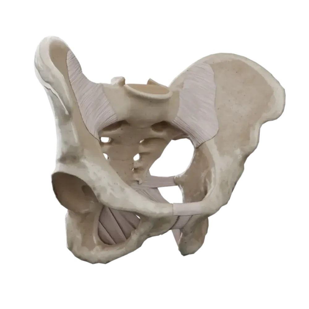
Pelvic joints anatomy test
Evaluate knowledge of pelvic joints anatomy. The test evaluates the structure of the sacroiliac joint, pubic symphysis, and ligamentous apparatus.
1. To which type of joints does the sacroiliac joint belong according to its classification?
-
Cylinder-shaped
The sacroiliac joint is a flat (tight) joint where movements are significantly limited by a strong ligamentous apparatus.
-
Ball-and-socket
The sacroiliac joint is a flat (tight) joint where movements are significantly limited by a strong ligamentous apparatus.
-
Flat
The sacroiliac joint is a flat (tight) joint where movements are significantly limited by a strong ligamentous apparatus.
-
Saddle-shaped
The sacroiliac joint is a flat (tight) joint where movements are significantly limited by a strong ligamentous apparatus.
-
I find it difficult to answer
The sacroiliac joint is a flat (tight) joint where movements are significantly limited by a strong ligamentous apparatus.
2. Which ligament closes the greater sciatic notch, transforming it into the greater sciatic foramen?
-
Sacrotuberous ligament
The sacrospinous ligament (lig. sacrospinale) extends between the sacrum and ischial spine, limiting the greater sciatic foramen from below.
-
Sacrospinous ligament
The sacrospinous ligament (lig. sacrospinale) extends between the sacrum and ischial spine, limiting the greater sciatic foramen from below.
-
Iliolumbar ligament
The sacrospinous ligament (lig. sacrospinale) extends between the sacrum and ischial spine, limiting the greater sciatic foramen from below.
-
Inguinal ligament
The sacrospinous ligament (lig. sacrospinale) extends between the sacrum and ischial spine, limiting the greater sciatic foramen from below.
-
I find it difficult to answer
The sacrospinous ligament (lig. sacrospinale) extends between the sacrum and ischial spine, limiting the greater sciatic foramen from below.
3. Between which anatomical structures does the iliolumbar ligament (lig. iliolumbale) extend?
-
Between the transverse processes of the IV-V lumbar vertebrae and the crest of the ilium
The iliolumbar ligament begins at the transverse processes of the lower lumbar vertebrae and attaches to the posterior part of the inner lip of the iliac crest.
-
Between the spinous processes of the lumbar vertebrae and the sacrum
The iliolumbar ligament begins at the transverse processes of the lower lumbar vertebrae and attaches to the posterior part of the inner lip of the iliac crest.
-
Between the twelfth rib and the ilium
The iliolumbar ligament begins at the transverse processes of the lower lumbar vertebrae and attaches to the posterior part of the inner lip of the iliac crest.
-
Between the promontory of the sacrum and the pubic symphysis
The iliolumbar ligament begins at the transverse processes of the lower lumbar vertebrae and attaches to the posterior part of the inner lip of the iliac crest.
-
I find it difficult to answer
The iliolumbar ligament begins at the transverse processes of the lower lumbar vertebrae and attaches to the posterior part of the inner lip of the iliac crest.
4. Which structure passes through the obturator canal (canalis obturatorius)?
-
Femoral nerve.
The obturator canal is formed by the obturator groove of the pubic bone and the superior edge of the obturator membrane; through it pass the homologous vessels and nerve.
-
Obturator vessels and nerve
The obturator canal is formed by the obturator groove of the pubic bone and the superior edge of the obturator membrane; through it pass the homologous vessels and nerve.
-
Sciatic nerve
The obturator canal is formed by the obturator groove of the pubic bone and the superior edge of the obturator membrane; through it pass the homologous vessels and nerve.
-
Internal pudendal artery
The obturator canal is formed by the obturator groove of the pubic bone and the superior edge of the obturator membrane; through it pass the homologous vessels and nerve.
-
I find it difficult to answer
The obturator canal is formed by the obturator groove of the pubic bone and the superior edge of the obturator membrane; through it pass the homologous vessels and nerve.
5. Which articular surfaces participate in forming the sacroiliac joint?
-
Auricular surfaces of the sacrum and ilium
The joint is formed by auricular surfaces (facies auriculares) of the sacrum and ilium, covered with fibrocartilage.
-
Bodies of sacral vertebrae and ischial bone
The joint is formed by auricular surfaces (facies auriculares) of the sacrum and ilium, covered with fibrocartilage.
-
Promontory of the sacrum and arcuate line of the ilium
The joint is formed by auricular surfaces (facies auriculares) of the sacrum and ilium, covered with fibrocartilage.
-
Sacral tuberosity and iliac tuberosity
The joint is formed by auricular surfaces (facies auriculares) of the sacrum and ilium, covered with fibrocartilage.
-
I find it difficult to answer
The joint is formed by auricular surfaces (facies auriculares) of the sacrum and ilium, covered with fibrocartilage.
6. Which of the listed ligaments is the strongest in the human body and is located within the sacroiliac joint?
-
Anterior sacroiliac ligament
The interosseous sacroiliac ligaments (ligg. sacroiliaca interossea) lie deeply between the tuberosities of the sacrum and ilium, being among the strongest in the body.
-
Interosseous sacroiliac ligament
The interosseous sacroiliac ligaments (ligg. sacroiliaca interossea) lie deeply between the tuberosities of the sacrum and ilium, being among the strongest in the body.
-
Sacrospinous ligament
The interosseous sacroiliac ligaments (ligg. sacroiliaca interossea) lie deeply between the tuberosities of the sacrum and ilium, being among the strongest in the body.
-
Posterior sacroiliac ligament
The interosseous sacroiliac ligaments (ligg. sacroiliaca interossea) lie deeply between the tuberosities of the sacrum and ilium, being among the strongest in the body.
-
I find it difficult to answer
The interosseous sacroiliac ligaments (ligg. sacroiliaca interossea) lie deeply between the tuberosities of the sacrum and ilium, being among the strongest in the body.
7. Which anatomical structure limits the lesser sciatic foramen from above?
-
Sacrotuberous ligament
The lesser sciatic foramen is bounded superiorly by the sacrospinous ligament, anteriorly by the ischial notch, and inferiorly and posteriorly by the sacrotuberous ligament.
-
Ischial tuberosity.
The lesser sciatic foramen is bounded superiorly by the sacrospinous ligament, anteriorly by the ischial notch, and inferiorly and posteriorly by the sacrotuberous ligament.
-
Sacrospinous ligament
The lesser sciatic foramen is bounded superiorly by the sacrospinous ligament, anteriorly by the ischial notch, and inferiorly and posteriorly by the sacrotuberous ligament.
-
Obturator membrane.
The lesser sciatic foramen is bounded superiorly by the sacrospinous ligament, anteriorly by the ischial notch, and inferiorly and posteriorly by the sacrotuberous ligament.
-
I find it difficult to answer
The lesser sciatic foramen is bounded superiorly by the sacrospinous ligament, anteriorly by the ischial notch, and inferiorly and posteriorly by the sacrotuberous ligament.
8. What type of joint is the pubic symphysis (symphysis pubica)?
-
Syndesmosis
The pubic symphysis represents a transitional form of joint (semi-joint or hemiarthrosis), as there is a narrow slit-like cavity without synovial membrane within the interpubic disc.
-
Hemiarthrosis (semi-joint)
The pubic symphysis represents a transitional form of joint (semi-joint or hemiarthrosis), as there is a narrow slit-like cavity without synovial membrane within the interpubic disc.
-
Diarthrosis
The pubic symphysis represents a transitional form of joint (semi-joint or hemiarthrosis), as there is a narrow slit-like cavity without synovial membrane within the interpubic disc.
-
Synchondrosis
The pubic symphysis represents a transitional form of joint (semi-joint or hemiarthrosis), as there is a narrow slit-like cavity without synovial membrane within the interpubic disc.
-
I find it difficult to answer
The pubic symphysis represents a transitional form of joint (semi-joint or hemiarthrosis), as there is a narrow slit-like cavity without synovial membrane within the interpubic disc.
9. What ligament strengthens the pubic symphysis at its lower edge?
-
Superior pubic ligament
The arcuate pubic ligament (lig. arcuatum pubis) is adjacent to the lower edge of the pubic symphysis, smoothing the subpubic angle.
-
Inguinal ligament
The arcuate pubic ligament (lig. arcuatum pubis) is adjacent to the lower edge of the pubic symphysis, smoothing the subpubic angle.
-
Arcuate pubic ligament
The arcuate pubic ligament (lig. arcuatum pubis) is adjacent to the lower edge of the pubic symphysis, smoothing the subpubic angle.
-
Pectineal ligament.
The arcuate pubic ligament (lig. arcuatum pubis) is adjacent to the lower edge of the pubic symphysis, smoothing the subpubic angle.
-
I find it difficult to answer
The arcuate pubic ligament (lig. arcuatum pubis) is adjacent to the lower edge of the pubic symphysis, smoothing the subpubic angle.
10. What is the name of the line that conventionally separates the greater and lesser pelvis?
-
Terminal line (linea terminalis).
The terminal line passes through the promontory of the sacrum, the arcuate lines of the iliac bones, the pubic crests, and the upper edge of the pubic symphysis.
-
Arcuate line (linea arcuata)
The terminal line passes through the promontory of the sacrum, the arcuate lines of the iliac bones, the pubic crests, and the upper edge of the pubic symphysis.
-
Linea alba
The terminal line passes through the promontory of the sacrum, the arcuate lines of the iliac bones, the pubic crests, and the upper edge of the pubic symphysis.
-
Intercrestal line (linea intercristalis)
The terminal line passes through the promontory of the sacrum, the arcuate lines of the iliac bones, the pubic crests, and the upper edge of the pubic symphysis.
-
I find it difficult to answer
The terminal line passes through the promontory of the sacrum, the arcuate lines of the iliac bones, the pubic crests, and the upper edge of the pubic symphysis.
11. What is the normal gynecological conjugate (conjugata vera) or true conjugate in women?
-
About 9 cm
The true (gynecological) conjugate is the distance from the sacral promontory to the most posterior point of the pubic symphysis, normally around 11 cm.
-
About 11 cm
The true (gynecological) conjugate is the distance from the sacral promontory to the most posterior point of the pubic symphysis, normally around 11 cm.
-
About 13 cm
The true (gynecological) conjugate is the distance from the sacral promontory to the most posterior point of the pubic symphysis, normally around 11 cm.
-
About 15 cm
The true (gynecological) conjugate is the distance from the sacral promontory to the most posterior point of the pubic symphysis, normally around 11 cm.
-
I find it difficult to answer
The true (gynecological) conjugate is the distance from the sacral promontory to the most posterior point of the pubic symphysis, normally around 11 cm.
12. What forms the lateral wall of the lesser pelvis?
-
Internal obturator muscle and piriformis muscle
The lateral walls of the lesser pelvis are formed by the pelvic bones below the linea terminalis, obturator membrane, sacrotuberous and sacrospinous ligaments.
-
Pelvic surface of the sacrum and coccyx
The lateral walls of the lesser pelvis are formed by the pelvic bones below the linea terminalis, obturator membrane, sacrotuberous and sacrospinous ligaments.
-
Bodies of the ischial bones and sacrotuberous ligaments
The lateral walls of the lesser pelvis are formed by the pelvic bones below the linea terminalis, obturator membrane, sacrotuberous and sacrospinous ligaments.
-
Internal surface of pelvic bones below the terminal line, obturator membrane, and ligaments
The lateral walls of the lesser pelvis are formed by the pelvic bones below the linea terminalis, obturator membrane, sacrotuberous and sacrospinous ligaments.
-
I find it difficult to answer
The lateral walls of the lesser pelvis are formed by the pelvic bones below the linea terminalis, obturator membrane, sacrotuberous and sacrospinous ligaments.
13. Which ligaments prevent the tilting of the sacral apex posteriorly (nutation) under spinal load?
-
Sacrotuberous and sacrospinous ligaments
The sacrotuberous and sacrospinous ligaments secure the lower part of the sacrum to the pelvic bone, preventing rotation of the sacrum around the transverse axis and displacement of its apex posteriorly.
-
Iliolumbar ligaments
The sacrotuberous and sacrospinous ligaments secure the lower part of the sacrum to the pelvic bone, preventing rotation of the sacrum around the transverse axis and displacement of its apex posteriorly.
-
Anterior longitudinal ligaments
The sacrotuberous and sacrospinous ligaments secure the lower part of the sacrum to the pelvic bone, preventing rotation of the sacrum around the transverse axis and displacement of its apex posteriorly.
-
Inguinal ligaments
The sacrotuberous and sacrospinous ligaments secure the lower part of the sacrum to the pelvic bone, preventing rotation of the sacrum around the transverse axis and displacement of its apex posteriorly.
-
I find it difficult to answer
The sacrotuberous and sacrospinous ligaments secure the lower part of the sacrum to the pelvic bone, preventing rotation of the sacrum around the transverse axis and displacement of its apex posteriorly.
14. Which opening is not related to the walls of the lesser pelvis?
-
Greater sciatic foramen
The femoral ring (anulus femoralis) is located under the inguinal ligament and pertains to the topography of the lower limb (thigh region) and not to the walls of the lesser pelvis.
-
Lesser sciatic foramen
The femoral ring (anulus femoralis) is located under the inguinal ligament and pertains to the topography of the lower limb (thigh region) and not to the walls of the lesser pelvis.
-
Obturator canal.
The femoral ring (anulus femoralis) is located under the inguinal ligament and pertains to the topography of the lower limb (thigh region) and not to the walls of the lesser pelvis.
-
Femoral ring
The femoral ring (anulus femoralis) is located under the inguinal ligament and pertains to the topography of the lower limb (thigh region) and not to the walls of the lesser pelvis.
-
I find it difficult to answer
The femoral ring (anulus femoralis) is located under the inguinal ligament and pertains to the topography of the lower limb (thigh region) and not to the walls of the lesser pelvis.
15. The anatomical substrate of the sacrococcygeal joint (articulatio sacrococcygea) is most often:
-
Typical synovial joint (diarthrosis)
The sacrococcygeal joint is a symphysis, within the intervertebral disc of which there is a small cavity, providing mobility to the coccyx.
-
Syndesmosis
The sacrococcygeal joint is a symphysis, within the intervertebral disc of which there is a small cavity, providing mobility to the coccyx.
-
Symphysis with an intervertebral disc and slit-like cavity
The sacrococcygeal joint is a symphysis, within the intervertebral disc of which there is a small cavity, providing mobility to the coccyx.
-
Synostosis in young people
The sacrococcygeal joint is a symphysis, within the intervertebral disc of which there is a small cavity, providing mobility to the coccyx.
-
I find it difficult to answer
The sacrococcygeal joint is a symphysis, within the intervertebral disc of which there is a small cavity, providing mobility to the coccyx.
16. What is the average inclination angle (inclinatio pelvis) of the pelvis in women in the vertical position?
-
35-40 degrees
The inclination angle of the pelvis is the angle between the horizontal plane and the plane of the superior pelvic aperture; in women, it is 55–60 degrees.
-
45-50 degrees
The inclination angle of the pelvis is the angle between the horizontal plane and the plane of the superior pelvic aperture; in women, it is 55–60 degrees.
-
55-60 degrees
The inclination angle of the pelvis is the angle between the horizontal plane and the plane of the superior pelvic aperture; in women, it is 55–60 degrees.
-
70-75 degrees
The inclination angle of the pelvis is the angle between the horizontal plane and the plane of the superior pelvic aperture; in women, it is 55–60 degrees.
-
I find it difficult to answer
The inclination angle of the pelvis is the angle between the horizontal plane and the plane of the superior pelvic aperture; in women, it is 55–60 degrees.
17. Which ligament passes along the anterior surface of the sacrum and coccyx, strengthening their connection?
-
Ventral sacrococcygeal ligament
The ventral sacrococcygeal ligament (lig. sacrococcygeum ventrale) is a continuation of the anterior longitudinal ligament of the spine and is located on the pelvic surface of the sacrum and coccyx.
-
Deep dorsal sacrococcygeal ligament
The ventral sacrococcygeal ligament (lig. sacrococcygeum ventrale) is a continuation of the anterior longitudinal ligament of the spine and is located on the pelvic surface of the sacrum and coccyx.
-
Superficial dorsal sacrococcygeal ligament
The ventral sacrococcygeal ligament (lig. sacrococcygeum ventrale) is a continuation of the anterior longitudinal ligament of the spine and is located on the pelvic surface of the sacrum and coccyx.
-
Lateral sacrococcygeal ligament
The ventral sacrococcygeal ligament (lig. sacrococcygeum ventrale) is a continuation of the anterior longitudinal ligament of the spine and is located on the pelvic surface of the sacrum and coccyx.
-
I find it difficult to answer
The ventral sacrococcygeal ligament (lig. sacrococcygeum ventrale) is a continuation of the anterior longitudinal ligament of the spine and is located on the pelvic surface of the sacrum and coccyx.
18. How is the anatomical conjugate (conjugata anatomica) measured?
-
From the sacral promontory to the lower edge of the pubic symphysis.
The anatomical conjugate (approximately 11.5 cm) is measured from the sacral promontory to the upper edge of the pubic symphysis, in contrast to the true conjugate.
-
From the sacral promontory to the upper edge of the pubic symphysis.
The anatomical conjugate (approximately 11.5 cm) is measured from the sacral promontory to the upper edge of the pubic symphysis, in contrast to the true conjugate.
-
From the sacral promontory to the most posteriorly prominent point of the symphysis.
The anatomical conjugate (approximately 11.5 cm) is measured from the sacral promontory to the upper edge of the pubic symphysis, in contrast to the true conjugate.
-
Between the arcuate lines of the ilium.
The anatomical conjugate (approximately 11.5 cm) is measured from the sacral promontory to the upper edge of the pubic symphysis, in contrast to the true conjugate.
-
I find it difficult to answer
The anatomical conjugate (approximately 11.5 cm) is measured from the sacral promontory to the upper edge of the pubic symphysis, in contrast to the true conjugate.
19. In which plane is the obturator membrane primarily situated?
-
In the sagittal.
The obturator membrane covers the obturator foramen of the pelvic bone, which is positioned obliquely, participating in the formation of the inferolateral wall of the pelvic cavity.
-
In the frontal.
The obturator membrane covers the obturator foramen of the pelvic bone, which is positioned obliquely, participating in the formation of the inferolateral wall of the pelvic cavity.
-
In the horizontal.
The obturator membrane covers the obturator foramen of the pelvic bone, which is positioned obliquely, participating in the formation of the inferolateral wall of the pelvic cavity.
-
At an angle, forming the inferolateral wall of the pelvis.
The obturator membrane covers the obturator foramen of the pelvic bone, which is positioned obliquely, participating in the formation of the inferolateral wall of the pelvic cavity.
-
I find it difficult to answer
The obturator membrane covers the obturator foramen of the pelvic bone, which is positioned obliquely, participating in the formation of the inferolateral wall of the pelvic cavity.
20. What movement does the sacrococcygeal joint provide during childbirth?
-
Forward displacement of the sacrum.
Due to the symphysial structure of the sacrococcygeal joint, it is possible for the coccyx to deviate backward during childbirth, increasing the anteroposterior diameter of the pelvic outlet.
-
Coccygeal backward deviation.
Due to the symphysial structure of the sacrococcygeal joint, it is possible for the coccyx to deviate backward during childbirth, increasing the anteroposterior diameter of the pelvic outlet.
-
Lateral flexion of the sacrum.
Due to the symphysial structure of the sacrococcygeal joint, it is possible for the coccyx to deviate backward during childbirth, increasing the anteroposterior diameter of the pelvic outlet.
-
Rotation of the coccyx around the vertical axis.
Due to the symphysial structure of the sacrococcygeal joint, it is possible for the coccyx to deviate backward during childbirth, increasing the anteroposterior diameter of the pelvic outlet.
-
I find it difficult to answer
Due to the symphysial structure of the sacrococcygeal joint, it is possible for the coccyx to deviate backward during childbirth, increasing the anteroposterior diameter of the pelvic outlet.
Retake this quiz?
Your current progress will be reset.