1/20


Anatomy test on the muscles of the abdomen and pelvis
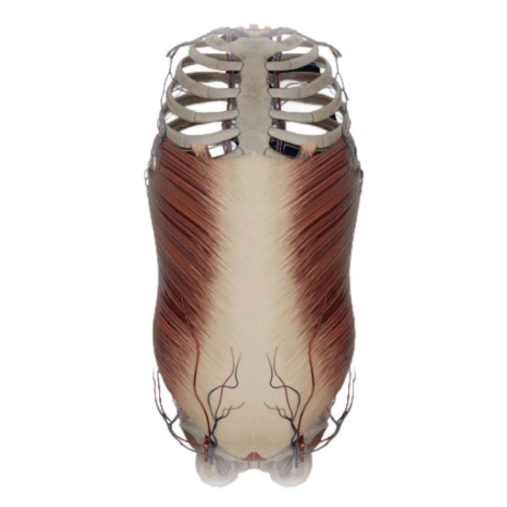
Evaluate your knowledge on the muscles of the abdomen and pelvis. The test covers their topography, fasciae, innervation, blood supply, and pelvic floor structure.
1. Which aponeurosis forms the anterior layer of the rectus sheath above the arcuate line (linea arcuata)?
-
Aponeuroses of internal oblique and transversus muscles
Above the linea arcuata, the anterior layer is formed by the aponeurosis of the external oblique and the superficial layer of the internal oblique muscle aponeurosis.
-
Only the aponeurosis of the transverse muscle
Above the linea arcuata, the anterior layer is formed by the aponeurosis of the external oblique and the superficial layer of the internal oblique muscle aponeurosis.
-
Aponeurosis of the external oblique and the anterior layer of the internal oblique muscle aponeurosis
Above the linea arcuata, the anterior layer is formed by the aponeurosis of the external oblique and the superficial layer of the internal oblique muscle aponeurosis.
-
Only the aponeurosis of the external oblique muscle
Above the linea arcuata, the anterior layer is formed by the aponeurosis of the external oblique and the superficial layer of the internal oblique muscle aponeurosis.
-
I find it difficult to answer
Above the linea arcuata, the anterior layer is formed by the aponeurosis of the external oblique and the superficial layer of the internal oblique muscle aponeurosis.
2. From which plexus is the quadratus lumborum muscle innervated?
-
Cervical plexus
The quadratus lumborum muscle receives motor innervation from the muscular branches of the lumbar plexus (Th12, L1-L4).
-
Brachial plexus
The quadratus lumborum muscle receives motor innervation from the muscular branches of the lumbar plexus (Th12, L1-L4).
-
Sacral plexus
The quadratus lumborum muscle receives motor innervation from the muscular branches of the lumbar plexus (Th12, L1-L4).
-
Lumbar plexus
The quadratus lumborum muscle receives motor innervation from the muscular branches of the lumbar plexus (Th12, L1-L4).
-
I find it difficult to answer
The quadratus lumborum muscle receives motor innervation from the muscular branches of the lumbar plexus (Th12, L1-L4).
3. The inguinal ligament (ligamentum inguinale) is the thickened inferolateral edge of the aponeurosis of which muscle?
-
External oblique muscle of the abdomen
The inguinal ligament represents the thickened lower edge of the aponeurosis of the external oblique muscle of the abdomen, stretched between the anterior superior iliac spine and the pubic tubercle.
-
Internal oblique muscle of the abdomen
The inguinal ligament represents the thickened lower edge of the aponeurosis of the external oblique muscle of the abdomen, stretched between the anterior superior iliac spine and the pubic tubercle.
-
Transversus abdominis muscle
The inguinal ligament represents the thickened lower edge of the aponeurosis of the external oblique muscle of the abdomen, stretched between the anterior superior iliac spine and the pubic tubercle.
-
Rectus abdominis muscle
The inguinal ligament represents the thickened lower edge of the aponeurosis of the external oblique muscle of the abdomen, stretched between the anterior superior iliac spine and the pubic tubercle.
-
I find it difficult to answer
The inguinal ligament represents the thickened lower edge of the aponeurosis of the external oblique muscle of the abdomen, stretched between the anterior superior iliac spine and the pubic tubercle.
4. Which muscles form the pelvic diaphragm (diaphragma pelvis)?
-
M. sphincter ani externus and m. transversus perinei profundus
The pelvic diaphragm is formed by the levator ani muscle and the coccygeus muscle, covered by the upper and lower fasciae of the pelvis.
-
M. levator ani and m. coccygeus
The pelvic diaphragm is formed by the levator ani muscle and the coccygeus muscle, covered by the upper and lower fasciae of the pelvis.
-
M. obturatorius internus and m. piriformis
The pelvic diaphragm is formed by the levator ani muscle and the coccygeus muscle, covered by the upper and lower fasciae of the pelvis.
-
M. ischiocavernosus and m. bulbospongiosus
The pelvic diaphragm is formed by the levator ani muscle and the coccygeus muscle, covered by the upper and lower fasciae of the pelvis.
-
I find it difficult to answer
The pelvic diaphragm is formed by the levator ani muscle and the coccygeus muscle, covered by the upper and lower fasciae of the pelvis.
5. Which muscle is part of the deep portion of the urogenital diaphragm (diaphragma urogenitale)?
-
M. transversus perinei superficialis
In the deep part of the urogenital region lies the deep transverse perineal muscle (m. transversus perinei profundus) and the urethral sphincter.
-
M. bulbospongiosus
In the deep part of the urogenital region lies the deep transverse perineal muscle (m. transversus perinei profundus) and the urethral sphincter.
-
M. transversus perinei profundus
In the deep part of the urogenital region lies the deep transverse perineal muscle (m. transversus perinei profundus) and the urethral sphincter.
-
M. ischiocavernosus
In the deep part of the urogenital region lies the deep transverse perineal muscle (m. transversus perinei profundus) and the urethral sphincter.
-
I find it difficult to answer
In the deep part of the urogenital region lies the deep transverse perineal muscle (m. transversus perinei profundus) and the urethral sphincter.
6. What is the anatomical substrate of the linea alba?
-
Interweaving of tendon fibers of the aponeuroses of broad abdominal muscles
The linea alba is formed along the median line by interweaving the tendinous bundles of the aponeuroses of the external, internal oblique, and transversus muscles from both sides.
-
Scarpa's fascia
The linea alba is formed along the median line by interweaving the tendinous bundles of the aponeuroses of the external, internal oblique, and transversus muscles from both sides.
-
Transverse fascia (fascia transversalis)
The linea alba is formed along the median line by interweaving the tendinous bundles of the aponeuroses of the external, internal oblique, and transversus muscles from both sides.
-
Tendinous intersections of the rectus abdominis muscle
The linea alba is formed along the median line by interweaving the tendinous bundles of the aponeuroses of the external, internal oblique, and transversus muscles from both sides.
-
I find it difficult to answer
The linea alba is formed along the median line by interweaving the tendinous bundles of the aponeuroses of the external, internal oblique, and transversus muscles from both sides.
7. From which muscles do the fibers of the cremaster muscle (m. cremaster) originate?
-
Rectus and pyramidalis abdominis muscles
The cremaster muscle is formed by bundles that separate from the internal oblique and transverse abdominal muscles as the testis descends.
-
External oblique and transverse abdominal muscles
The cremaster muscle is formed by bundles that separate from the internal oblique and transverse abdominal muscles as the testis descends.
-
Quadratus lumborum muscle and external oblique muscle
The cremaster muscle is formed by bundles that separate from the internal oblique and transverse abdominal muscles as the testis descends.
-
Internal oblique and transverse abdominal muscles
The cremaster muscle is formed by bundles that separate from the internal oblique and transverse abdominal muscles as the testis descends.
-
I find it difficult to answer
The cremaster muscle is formed by bundles that separate from the internal oblique and transverse abdominal muscles as the testis descends.
8. Which anatomical structure is located in the gap between m. psoas major and m. iliacus (lacuna musculorum)?
-
Femoral artery
Through the muscular space under the inguinal ligament, the iliopsoas muscle and femoral nerve (n. femoralis) exit to the thigh.
-
Femoral nerve
Through the muscular space under the inguinal ligament, the iliopsoas muscle and femoral nerve (n. femoralis) exit to the thigh.
-
Femoral vein
Through the muscular space under the inguinal ligament, the iliopsoas muscle and femoral nerve (n. femoralis) exit to the thigh.
-
Obturator nerve
Through the muscular space under the inguinal ligament, the iliopsoas muscle and femoral nerve (n. femoralis) exit to the thigh.
-
I find it difficult to answer
Through the muscular space under the inguinal ligament, the iliopsoas muscle and femoral nerve (n. femoralis) exit to the thigh.
9. Which of the listed muscles lines the lateral wall of the lesser pelvis?
-
M. obturatorius internus
The internal obturator muscle (m. obturatorius internus) originates from the internal surface of the obturator membrane and pelvic bones, lining its lateral wall.
-
M. piriformis
The internal obturator muscle (m. obturatorius internus) originates from the internal surface of the obturator membrane and pelvic bones, lining its lateral wall.
-
M. psoas minor
The internal obturator muscle (m. obturatorius internus) originates from the internal surface of the obturator membrane and pelvic bones, lining its lateral wall.
-
M. iliacus
The internal obturator muscle (m. obturatorius internus) originates from the internal surface of the obturator membrane and pelvic bones, lining its lateral wall.
-
I find it difficult to answer
The internal obturator muscle (m. obturatorius internus) originates from the internal surface of the obturator membrane and pelvic bones, lining its lateral wall.
10. Which abdominal muscle has characteristic tendon intersections (intersectiones tendineae)?
-
External oblique muscle of the abdomen
The rectus abdominis muscle (m. rectus abdominis) is interrupted along its course by 3-4 tendinous intersections, which blend with the anterior wall of its sheath.
-
Quadratus lumborum muscle
The rectus abdominis muscle (m. rectus abdominis) is interrupted along its course by 3-4 tendinous intersections, which blend with the anterior wall of its sheath.
-
Transversus abdominis muscle
The rectus abdominis muscle (m. rectus abdominis) is interrupted along its course by 3-4 tendinous intersections, which blend with the anterior wall of its sheath.
-
Rectus abdominis muscle
The rectus abdominis muscle (m. rectus abdominis) is interrupted along its course by 3-4 tendinous intersections, which blend with the anterior wall of its sheath.
-
I find it difficult to answer
The rectus abdominis muscle (m. rectus abdominis) is interrupted along its course by 3-4 tendinous intersections, which blend with the anterior wall of its sheath.
11. Muscle bundles of which part of the diaphragm delimit the esophageal hiatus (hiatus esophageus)?
-
Sternal part
The esophageal hiatus is formed by crossing medial crura of the lumbar part (pars lumbalis) of the diaphragm.
-
Lumbar part
The esophageal hiatus is formed by crossing medial crura of the lumbar part (pars lumbalis) of the diaphragm.
-
Costal part
The esophageal hiatus is formed by crossing medial crura of the lumbar part (pars lumbalis) of the diaphragm.
-
Tendinous center
The esophageal hiatus is formed by crossing medial crura of the lumbar part (pars lumbalis) of the diaphragm.
-
I find it difficult to answer
The esophageal hiatus is formed by crossing medial crura of the lumbar part (pars lumbalis) of the diaphragm.
12. Which artery supplies the rectus abdominis muscle, passing through its sheath from below upwards?
-
A. epigastrica superior
The inferior epigastric artery (a. epigastrica inferior) arises from the external iliac artery, penetrates the sheath of the rectus abdominis muscle posteriorly, and ascends.
-
A. thoracica interna
The inferior epigastric artery (a. epigastrica inferior) arises from the external iliac artery, penetrates the sheath of the rectus abdominis muscle posteriorly, and ascends.
-
A. epigastrica inferior
The inferior epigastric artery (a. epigastrica inferior) arises from the external iliac artery, penetrates the sheath of the rectus abdominis muscle posteriorly, and ascends.
-
A. circumflexa ilium profunda
The inferior epigastric artery (a. epigastrica inferior) arises from the external iliac artery, penetrates the sheath of the rectus abdominis muscle posteriorly, and ascends.
-
I find it difficult to answer
The inferior epigastric artery (a. epigastrica inferior) arises from the external iliac artery, penetrates the sheath of the rectus abdominis muscle posteriorly, and ascends.
13. Through which foramen does the piriformis muscle (m. piriformis) exit the pelvic cavity?
-
Foramen ischiadicum majus
The piriformis muscle originates on the pelvic surface of the sacrum and exits the pelvis through the greater sciatic foramen (foramen ischiadicum majus).
-
Foramen ischiadicum minus
The piriformis muscle originates on the pelvic surface of the sacrum and exits the pelvis through the greater sciatic foramen (foramen ischiadicum majus).
-
Canalis obturatorius
The piriformis muscle originates on the pelvic surface of the sacrum and exits the pelvis through the greater sciatic foramen (foramen ischiadicum majus).
-
Foramen suprapiriforme
The piriformis muscle originates on the pelvic surface of the sacrum and exits the pelvis through the greater sciatic foramen (foramen ischiadicum majus).
-
I find it difficult to answer
The piriformis muscle originates on the pelvic surface of the sacrum and exits the pelvis through the greater sciatic foramen (foramen ischiadicum majus).
14. What is the anterior boundary of the perineum (perineum)?
-
Apex of coccyx
The perineum has a rhomboid shape, with its anterior angle (boundary) being the inferior margin of the pubic symphysis.
-
Pubic symphysis
The perineum has a rhomboid shape, with its anterior angle (boundary) being the inferior margin of the pubic symphysis.
-
Ischial tuberosities
The perineum has a rhomboid shape, with its anterior angle (boundary) being the inferior margin of the pubic symphysis.
-
Sacrotuberous ligaments
The perineum has a rhomboid shape, with its anterior angle (boundary) being the inferior margin of the pubic symphysis.
-
I find it difficult to answer
The perineum has a rhomboid shape, with its anterior angle (boundary) being the inferior margin of the pubic symphysis.
15. To which group of abdominal muscles does the pyramidalis muscle (m. pyramidalis) belong?
-
To the muscles of the posterior abdominal wall
The pyramidalis muscle is located in the lower part of the anterior abdominal wall, within the sheath of the rectus abdominis muscle, and stretches the linea alba.
-
To the muscles of the lateral abdominal wall
The pyramidalis muscle is located in the lower part of the anterior abdominal wall, within the sheath of the rectus abdominis muscle, and stretches the linea alba.
-
Rudimentary muscle of the pelvic floor
The pyramidalis muscle is located in the lower part of the anterior abdominal wall, within the sheath of the rectus abdominis muscle, and stretches the linea alba.
-
To the muscles of the anterior abdominal wall
The pyramidalis muscle is located in the lower part of the anterior abdominal wall, within the sheath of the rectus abdominis muscle, and stretches the linea alba.
-
I find it difficult to answer
The pyramidalis muscle is located in the lower part of the anterior abdominal wall, within the sheath of the rectus abdominis muscle, and stretches the linea alba.
16. Which ligament, formed by the periosteum of the pubic bone, is the continuation of the lacunar ligament along the crest of the pubic bone?
-
Pectineal ligament (ligamentum pectineum)
The pectineal ligament (Cooper's ligament) stretches from the lacunar ligament laterally along the crest of the pubic bone (pecten ossis pubis).
-
Inguinal ligament (ligamentum inguinale)
The pectineal ligament (Cooper's ligament) stretches from the lacunar ligament laterally along the crest of the pubic bone (pecten ossis pubis).
-
Reflected ligament (ligamentum reflexum)
The pectineal ligament (Cooper's ligament) stretches from the lacunar ligament laterally along the crest of the pubic bone (pecten ossis pubis).
-
Iliopectineal arch (arcus iliopectineus)
The pectineal ligament (Cooper's ligament) stretches from the lacunar ligament laterally along the crest of the pubic bone (pecten ossis pubis).
-
I find it difficult to answer
The pectineal ligament (Cooper's ligament) stretches from the lacunar ligament laterally along the crest of the pubic bone (pecten ossis pubis).
17. Which of the following muscles is located in the superficial perineal space in males?
-
M. levator ani
The bulbospongiosus muscle (m. bulbospongiosus) surrounds the bulb of the penis and belongs to the superficial muscles of the urogenital region.
-
M. coccygeus
The bulbospongiosus muscle (m. bulbospongiosus) surrounds the bulb of the penis and belongs to the superficial muscles of the urogenital region.
-
M. bulbospongiosus
The bulbospongiosus muscle (m. bulbospongiosus) surrounds the bulb of the penis and belongs to the superficial muscles of the urogenital region.
-
M. transversus perinei profundus
The bulbospongiosus muscle (m. bulbospongiosus) surrounds the bulb of the penis and belongs to the superficial muscles of the urogenital region.
-
I find it difficult to answer
The bulbospongiosus muscle (m. bulbospongiosus) surrounds the bulb of the penis and belongs to the superficial muscles of the urogenital region.
18. To which anatomical structure of the femur does the iliopsoas muscle (m. iliopsoas) attach?
-
To the greater trochanter
The iliopsoas muscle, exiting to the thigh through the muscular lacuna, attaches to the lesser trochanter (trochanter minor) of the femur.
-
To the lesser trochanter
The iliopsoas muscle, exiting to the thigh through the muscular lacuna, attaches to the lesser trochanter (trochanter minor) of the femur.
-
To the intertrochanteric line
The iliopsoas muscle, exiting to the thigh through the muscular lacuna, attaches to the lesser trochanter (trochanter minor) of the femur.
-
To the gluteal tuberosity
The iliopsoas muscle, exiting to the thigh through the muscular lacuna, attaches to the lesser trochanter (trochanter minor) of the femur.
-
I find it difficult to answer
The iliopsoas muscle, exiting to the thigh through the muscular lacuna, attaches to the lesser trochanter (trochanter minor) of the femur.
19. In the splitting of the fascia of which muscle is the pudendal canal (Alcock's canal) formed?
-
Obturator externus muscle
The pudendal canal is located on the lateral wall of the ischioanal fossa and is formed by the splitting of the fascia of the obturator internus muscle (fascia obturatoria).
-
Piriformis muscle
The pudendal canal is located on the lateral wall of the ischioanal fossa and is formed by the splitting of the fascia of the obturator internus muscle (fascia obturatoria).
-
Levator ani muscle
The pudendal canal is located on the lateral wall of the ischioanal fossa and is formed by the splitting of the fascia of the obturator internus muscle (fascia obturatoria).
-
Obturator internus muscle
The pudendal canal is located on the lateral wall of the ischioanal fossa and is formed by the splitting of the fascia of the obturator internus muscle (fascia obturatoria).
-
I find it difficult to answer
The pudendal canal is located on the lateral wall of the ischioanal fossa and is formed by the splitting of the fascia of the obturator internus muscle (fascia obturatoria).
20. Which nerves predominantly provide motor innervation to the transversus abdominis muscle?
-
Only by the ilioinguinal nerve
The broad muscles of the abdomen are innervated by the lower six intercostal nerves (nn. intercostales VII-XII), as well as by branches of the lumbar plexus: n. iliohypogastricus and n. ilioinguinalis.
-
By branches of the sacral plexus
The broad muscles of the abdomen are innervated by the lower six intercostal nerves (nn. intercostales VII-XII), as well as by branches of the lumbar plexus: n. iliohypogastricus and n. ilioinguinalis.
-
By intercostal nerves (VII-XII), iliohypogastric and ilioinguinal nerves
The broad muscles of the abdomen are innervated by the lower six intercostal nerves (nn. intercostales VII-XII), as well as by branches of the lumbar plexus: n. iliohypogastricus and n. ilioinguinalis.
-
By branches of the vagus nerve
The broad muscles of the abdomen are innervated by the lower six intercostal nerves (nn. intercostales VII-XII), as well as by branches of the lumbar plexus: n. iliohypogastricus and n. ilioinguinalis.
-
I find it difficult to answer
The broad muscles of the abdomen are innervated by the lower six intercostal nerves (nn. intercostales VII-XII), as well as by branches of the lumbar plexus: n. iliohypogastricus and n. ilioinguinalis.
Retake this quiz?
Your current progress will be reset.